


Tuberculosis outside the lung usually results from hematogenous dissemination. Sometimes infection directly extends from an adjacent organ. Symptoms vary by site but generally include fever, malaise, and weight loss. Diagnosis is most often by sputum smear and culture and, increasingly, by rapid molecular-based diagnostic tests. Treatment is with multiple antimicrobial drugs given for at least 6 months.
Tuberculosis (TB) properly refers only to disease caused by Mycobacterium tuberculosis (for which humans are the main reservoir). Although the lungs are the initial site of infection, disease can spread to many organs. (For details on the organism, pathophysiology, and pulmonary disease, see Tuberculosis.)
Miliary tuberculosis
Also known as generalized hematogenous TB, miliary TB occurs when a tuberculous lesion erodes into a blood vessel, disseminating millions of tubercle bacilli into the bloodstream and throughout the body. Uncontrolled massive dissemination can occur during primary infection or after reactivation of a latent focus. The lungs and bone marrow are most often affected, but any site may be involved.
Miliary TB is most common among
Children < 4 years old
Immunocompromised people
Older people
Symptoms of miliary TB include fever, chills, weakness, malaise, and often progressive dyspnea. Intermittent dissemination of tubercle bacilli may lead to a prolonged fever of unknown origin (FUO). Bone marrow involvement may cause anemia, thrombocytopenia, or a leukemoid reaction.
Genitourinary tuberculosis
Infection of the kidneys may manifest as pyelonephritis (eg, fever, back pain, pyuria) without the usual urinary pathogens on routine culture (sterile pyuria). Infection commonly spreads to the bladder and, in men, to the prostate, seminal vesicles, or epididymis, causing an enlarging scrotal mass. Infection may spread to the perinephric space and down the psoas muscle, sometimes causing an abscess on the anterior thigh.
Salpingo-oophoritis can occur after menarche, when the fallopian tubes become vascular. Symptoms include chronic pelvic pain and sterility or ectopic pregnancy due to tubal scarring.
Meningeal tuberculosis (TB meningitis)
Meningitis often occurs in the absence of infection at other extrapulmonary sites. In the US, it is most common among the elderly and immunocompromised, but in areas where TB is common among children, TB meningitis usually occurs between birth and 5 years. At any age, meningitis is the most serious form of TB and has high morbidity and mortality. It is the one form of TB shown to be prevented in childhood by vaccination with BCG.
Symptoms are low-grade fever, unremitting headache, nausea, and drowsiness, which may progress to stupor and coma. Kernig and Brudzinski signs may be positive. Because the early signs are non-specific, it is important to consider the diagnosis early in any patient with known TB exposure, infection, or disease, including past TB, and in all persons with compatible symptoms from high TB-burden locations. Stages are
1: Clear sensorium with abnormal CSF
2: Drowsiness or stupor with focal neurologic signs
3: Coma
Stroke may result from thrombosis of a major cerebral vessel. Focal neurologic symptoms suggest a tuberculoma.
Peritoneal tuberculosis (TB peritonitis)
Peritoneal infection represents seeding from abdominal lymph nodes or from salpingo-oophoritis. Peritonitis is particularly common among people with alcohol use disorder who have cirrhosis.
Symptoms may be mild, with fatigue, abdominal pain, and tenderness, or severe enough to mimic acute abdomen.
Pericardial tuberculosis (TB pericarditis)
Pericardial infection may develop from foci in mediastinal lymph nodes or from pleural TB. In some high-incidence parts of the world, TB pericarditis is a common cause of heart failure.
Patients may have a pericardial friction rub, pleuritic and positional chest pain, or fever. Pericardial tamponade may occur, causing dyspnea, neck vein distention, paradoxical pulse, muffled heart sounds, and possibly hypotension.
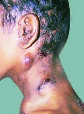
Tuberculous lymphadenitis
Tuberculous lymphadenitis (scrofula) typically involves the lymph nodes in the posterior cervical and supraclavicular chains. Infection in these areas is thought to be due to contiguous spread from intrathoracic lymphatics or from infection in the tonsils and adenoids. Mediastinal lymph nodes are also commonly enlarged as a part of primary pulmonary disease.
Cervical tuberculous lymphadenitis is characterized by progressive swelling of the affected nodes. In advanced cases, nodes may become inflamed and tender; the overlying skin may break down, resulting in a draining fistula.
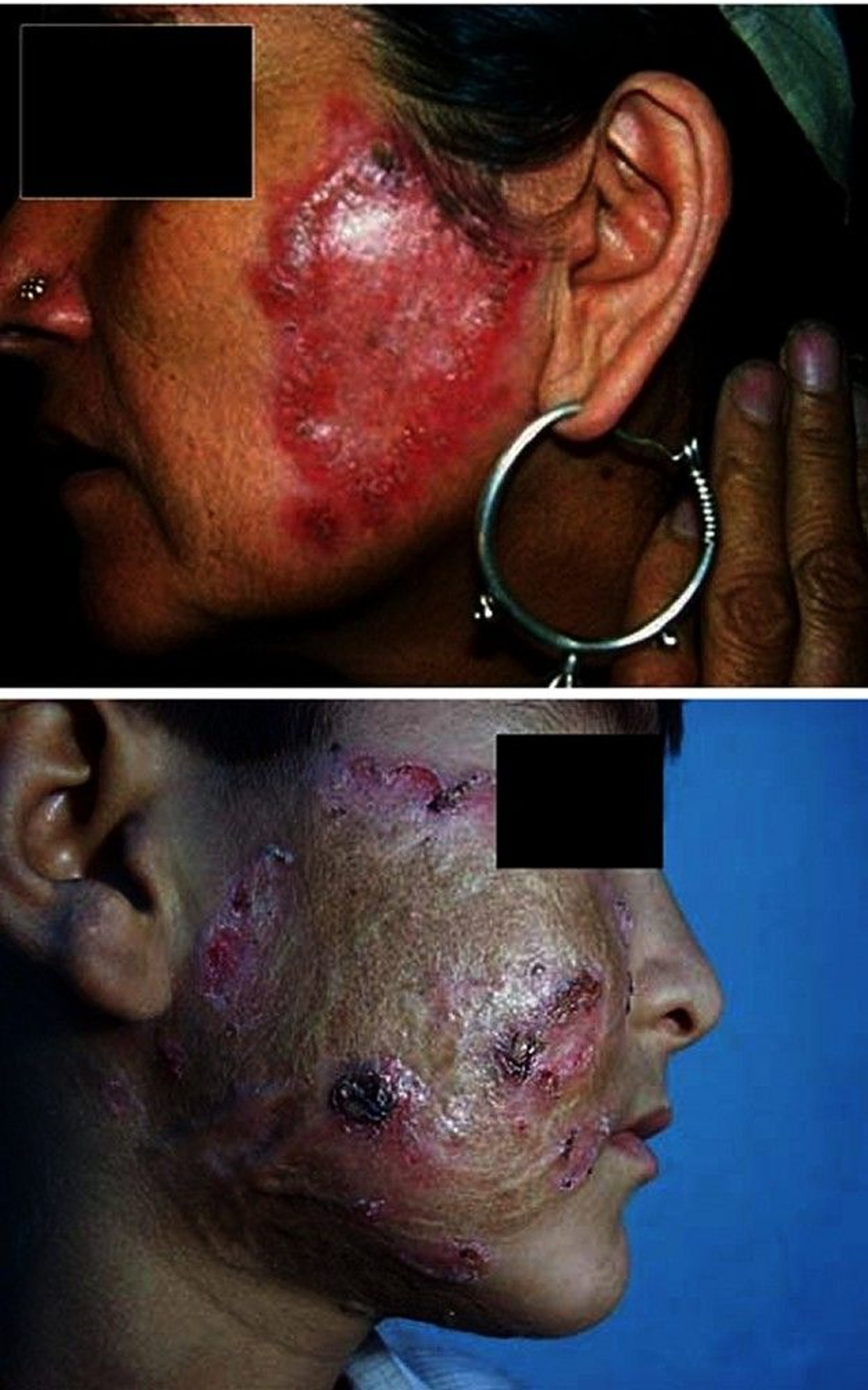
Cutaneous tuberculosis
Cutaneous tuberculosis (scrofuloderma) results from direct extension of an underlying TB focus (eg, a regional lymph node, an infected bone or joint) to the overlying skin, forming ulcers and sinus tracts.

Lupus vulgaris results from hematogenous or lymphogenous dissemination to the skin from an extracutaneous focus in a sensitized patient.

© Springer Science+Business Media
Tuberculosis verrucosa cutis (prosector's wart) occurs after exogenous direct inoculation of the mycobacteria into the skin of a previously sensitized patient who has moderate to high immunity against the bacilli.
Rarely, TB develops on abraded skin in patients with cavitary pulmonary TB.
Tuberculosis of bones and joints
Weight-bearing joints are most commonly involved, but bones of the wrist, hand, and elbow may also be affected, especially after injury.
Pott disease is spinal infection, which begins in a vertebral body and often spreads to adjacent vertebrae, with narrowing of the disk space between them. Untreated, the vertebrae may collapse, possibly impinging on the spinal cord. Symptoms include progressive or constant pain in involved bones and chronic or subacute arthritis (usually monoarticular). In Pott disease, spinal cord compression causes neurologic deficits, including paraplegia; paravertebral swelling may result from an abscess.
Gastrointestinal tuberculosis
Because the entire gastrointestinal (GI) mucosa resists TB invasion, infection requires prolonged exposure and enormous inocula. It is very unusual in countries where bovine TB is rare (eg, because of milk pasteurization and routine TB testing of cattle).
Ulcers of the mouth and oropharynx may develop from eating M. bovis–contaminated dairy products; primary lesions may also occur in the small bowel. Intestinal invasion generally causes hyperplasia and an inflammatory bowel syndrome with pain, diarrhea, obstruction, and hematochezia. It may also mimic appendicitis. Ulceration and fistulas are possible.
Tuberculosis of the liver
Liver infection is common in patients with advanced pulmonary TB and widely disseminated or miliary TB. However, the liver generally heals without sequelae when the principal infection is treated. TB in the liver occasionally spreads to the gallbladder, leading to obstructive jaundice.
Other sites of TB infection
TB may infect the wall of a blood vessel and has even ruptured the aorta. Adrenal involvement, leading to Addison disease, was common but now is rare. Tubercle bacilli may spread to tendon sheaths (tuberculous tenosynovitis) by direct extension from adjacent lesions in bone or hematogenously from any infected organ.
Diagnosis of Extrapulmonary TB
Acid-fast staining, microscopic analysis, and mycobacterial culture of fluid and tissue samples, and, when available, nucleic acid amplification testing (NAAT)
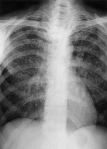
Chest x-ray
Tuberculin skin test (TST) or interferon-gamma release assay (IGRA)
Testing is similar to that for pulmonary TB (see Diagnosis of TB), including chest x-ray, TST or IGRA, and microscopic analysis (with appropriate staining) and mycobacterial cultures of affected body fluids (cerebrospinal fluid, urine, or pleural, pericardial, or joint fluid) and tissue for mycobacteria. Blood culture results are positive in about 50% of patients with disseminated TB; such patients are often immunocompromised, often by HIV infection. However, cultures and smears of body fluids and tissues are often negative because few organisms are present; in such cases, NAATs may be helpful.
NAATs can be done on fresh fluid or biopsy samples and on fixed tissue (eg, if TB was not suspected during a surgical procedure and cultures were not done). NAATs typically are not approved for extrapulmonary TB diagnosis but are commonly used in hopes of an early diagnosis for medical care and public health reasons, pending culture. Although a positive NAAT result almost always supports a TB diagnosis, a negative result does not rule out TB in most cases because the negative predictive value is generally unknown and may depend on specimen processing and other factors that are not standardized.
Typically, lymphocytosis is present in body fluids. A very suggestive finding in the cerebrospinal fluid is a glucose level < 50% of that in serum and an elevated protein level.
If all tests are negative and miliary TB is still a concern, biopsies of the bone marrow and the liver are done. If TB is highly suspected based on other features (eg, granuloma seen on biopsy, positive TST or IGRA result plus unexplained lymphocytosis in pleural fluid or cerebrospinal fluid), treatment should usually proceed despite inability to demonstrate TB organisms.
Chest x-ray and other imaging can also provide helpful diagnostic information. Chest x-ray may show signs of primary or active TB; in miliary TB, it shows thousands of 2- to 3-mm interstitial nodules evenly distributed through both lungs.

Other imaging tests are done based on clinical findings. Abdominal or genitourinary involvement usually requires CT or ultrasonography; renal lesions are often visible. Bone and joint involvement requires CT or MRI; MRI is preferable for spinal disease.
TST and IGRA may initially be negative, but a repeat test in a few weeks is likely to be positive. If it is not, the diagnosis of TB should be questioned or causes of anergy (no reaction to any skin test) should be sought.
Treatment of Extrapulmonary TB
Antibiotics
Sometimes corticosteroids
Sometimes surgery
Drug treatment is the most important modality and follows standard regimens and principles (see First-line drugs for TB). Six to 9 months of therapy is probably adequate for most sites except the meninges, which require treatment for 9 to 12 months.
Drug resistance is a major concern; it is increased by poor adherence, use of too few drugs, and inadequate susceptibility testing.
Corticosteroids are often used for TB meningitis, but the Centers for Disease Control and Prevention (CDC) no longer recommends corticosteroids for TB pericarditis that is not constrictive; however, corticosteroids may prevent constriction in patients who are at risk. Corticosteroids may be used for meningitis and TB pericarditis (even when not constrictive) in patients with immune reconstitution inflammatory syndrome.
Surgery is required for the following:
To drain empyema, cardiac tamponade, or central nervous system abscess
To close bronchopleural fistulas
To resect infected bowel
To decompress spinal cord encroachment
Surgical debridement is sometimes needed in Pott disease to correct spinal deformities or to relieve cord compression if there are neurologic deficits or pain persists; fixation of the vertebral column by bone graft is required in only the most advanced cases. Surgery is usually not necessary for TB lymphadenitis except for diagnostic purposes.
Key Points
Tuberculosis can spread from the lungs through the bloodstream to many sites.
Symptoms depend on the affected organ but typically include fever, malaise, and weight loss.
Diagnose based on identification of bacilli in infected fluid or tissue by microscopic examination and culture and/or nucleic acid amplification tests.
Treat with multiple drugs for several months and sometimes with surgery.
Drug resistance is a major concern.


