


Photosensitivity is a cutaneous overreaction to sunlight. It can be related to photoallergy or phototoxicity and may be idiopathic or occur after exposure to certain toxic or allergenic substances or chemicals. It can also sometimes be a feature of systemic disorders (eg, systemic lupus erythematosus, porphyria, pellagra, xeroderma pigmentosum). Diagnosis is clinical. Treatment varies by type.
In addition to the acute effects of sunlight and the chronic effects of sunlight, a variety of less common reactions may occur after sun exposure. Unless the cause is obvious, patients with pronounced photosensitivity should be evaluated for systemic or cutaneous disorders associated with light sensitivity such as systemic lupus erythematosus (SLE), dermatomyositis, and porphyria.
(See also Overview of Effects of Sunlight.)
Solar urticaria
In certain patients, urticaria develops at a site of sun exposure within a few minutes. Lesions generally resolve within 24 hours. Rarely, if large areas are involved, syncope, dizziness, wheezing, and other systemic symptoms may develop. Etiology is unclear but may involve endogenous skin constituents functioning as photoallergens, leading to mast cell degranulation as in other types of urticaria. Solar urticaria can be distinguished from other types of urticaria in that wheals in solar urticaria occur only on exposed skin after ultraviolet (UV) light exposure.

Solar urticaria can be classified based on the component of the UV spectrum (UVA, UVB, and visible light) that causes lesions. If necessary, patients can be tested by exposing part of the skin to natural light or artificial light at particular wavelengths (phototesting).
Chemical photosensitivity
Over 100 substances, ingested or applied topically, are known to predispose to cutaneous reactions after sun exposure. A limited number are responsible for most reactions (see table Some Medications or Substances That Cause Cutaneous Photosensitivity). Reactions are divided into phototoxicity and photoallergy. Phototesting can help confirm the diagnosis. Treatment for chemical photosensitivity is topical corticosteroids and avoidance of the causative substance.

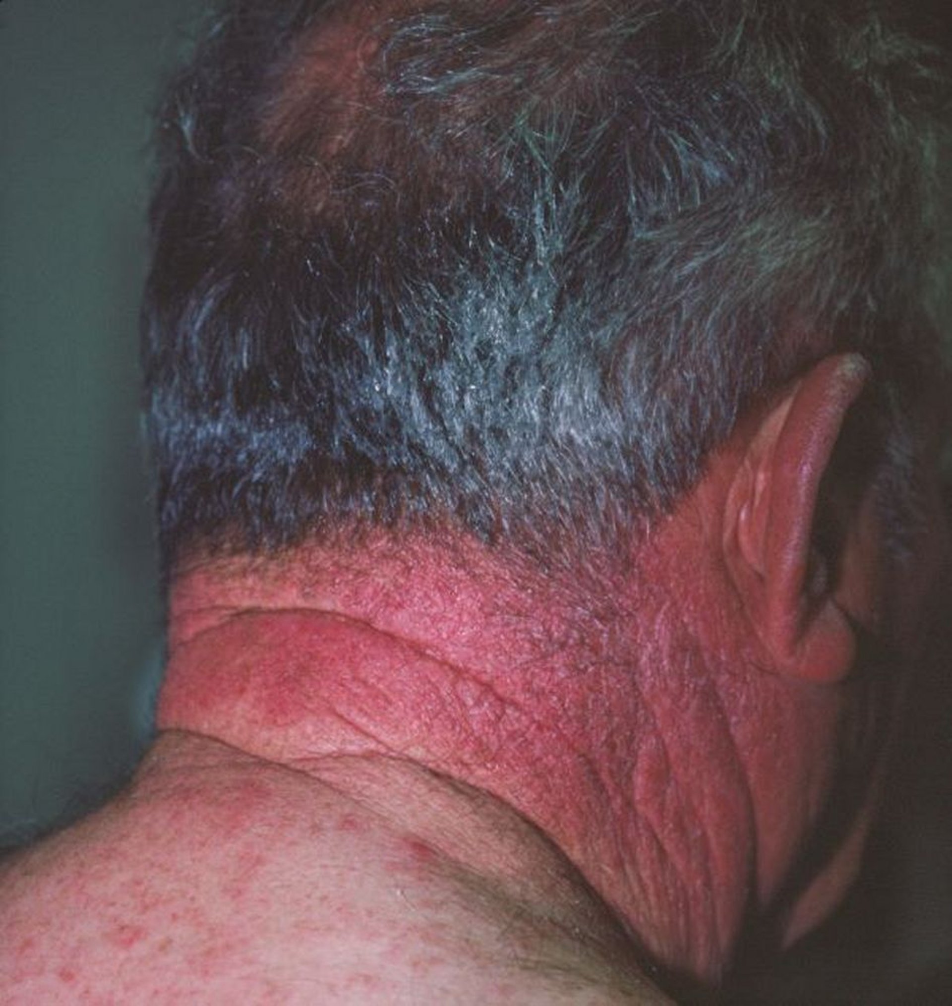
Image courtesy of Karen McKoy, MD.
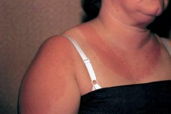
In phototoxicity, light-absorbing compounds directly generate free radicals and inflammatory mediators, causing tissue damage manifesting as pain and erythema (like sunburn
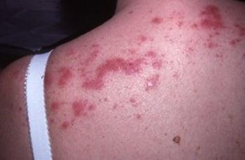
Photoallergy is a type IV (cell-mediated) immune response. Light absorption causes structural changes in the drug or substance, allowing it to bind to tissue protein and function as a hapten, making the complex allergenic. Prior exposure to the allergen is required. The reaction is usually eczematous, with erythema, scaling, pruritus, and sometimes vesicles. Typical causes of photoallergic reactions include aftershave lotions, sunscreens, and sulfonamides. Photoallergy occurs less often than phototoxicity, and the reaction may extend to non–sun-exposed skin.
Polymorphous light eruption
Polymorphous light eruption is a common photosensitive reaction to UV and sometimes visible light. It does not seem to be associated with systemic disease or drugs. A positive family history in some patients suggests a genetic risk factor.
Eruptions appear on sun-exposed areas, usually 30 minutes to several hours after exposure; however, sometimes eruptions do not appear for up to several days. Lesions are pruritic, erythematous, and often papular but may be papulovesicular or plaquelike. They are more common among women and people from northern climates when first exposed to spring or summer sun than among those exposed to sun year-round. Lesions often subside within several days to weeks.

Diagnosis of polymorphous light eruption is made by history, skin findings, and exclusion of other sun-sensitivity disorders. Diagnosis sometimes requires reproduction of the lesions with phototesting when the patient is not using any potentially photosensitizing drugs.
Often, lesions are self-limited and spontaneously improve as summer progresses. Preventive measures include using a broad-spectrum sunscreen and moderating sun exposure. More severely affected patients may benefit from desensitization in early spring by graduated exposure to UV light with low-dose psoralen plus ultraviolet A (PUVA― see Phototherapy
Some evidence suggests that antioxidants such as the dietary supplement Polypodium leucotomos, a natural tropical fern extract, may help prevent polymorphous light eruption, but further studies are needed (1).
General reference
1. Nestor MS, Berman B, Swenson N: Safety and efficacy of oral Polypodium leucotomos extract in healthy adult subjects. J Clin Aesthet Dermatol 8(2):19–23, 2015. PMID: 25741399


