


Hydrocephalus is accumulation of excessive amounts of cerebrospinal fluid, causing cerebral ventricular enlargement and/or increased intracranial pressure. Manifestations can include enlarged head, bulging fontanelle, irritability, lethargy, vomiting, and seizures. Diagnosis is by ultrasonography in neonates and young infants with an open fontanelle and by CT or MRI in older infants and children. Treatment ranges from observation to surgical intervention, depending on severity and progression of symptoms.

STEVE ALLEN/SCIENCE PHOTO LIBRARY
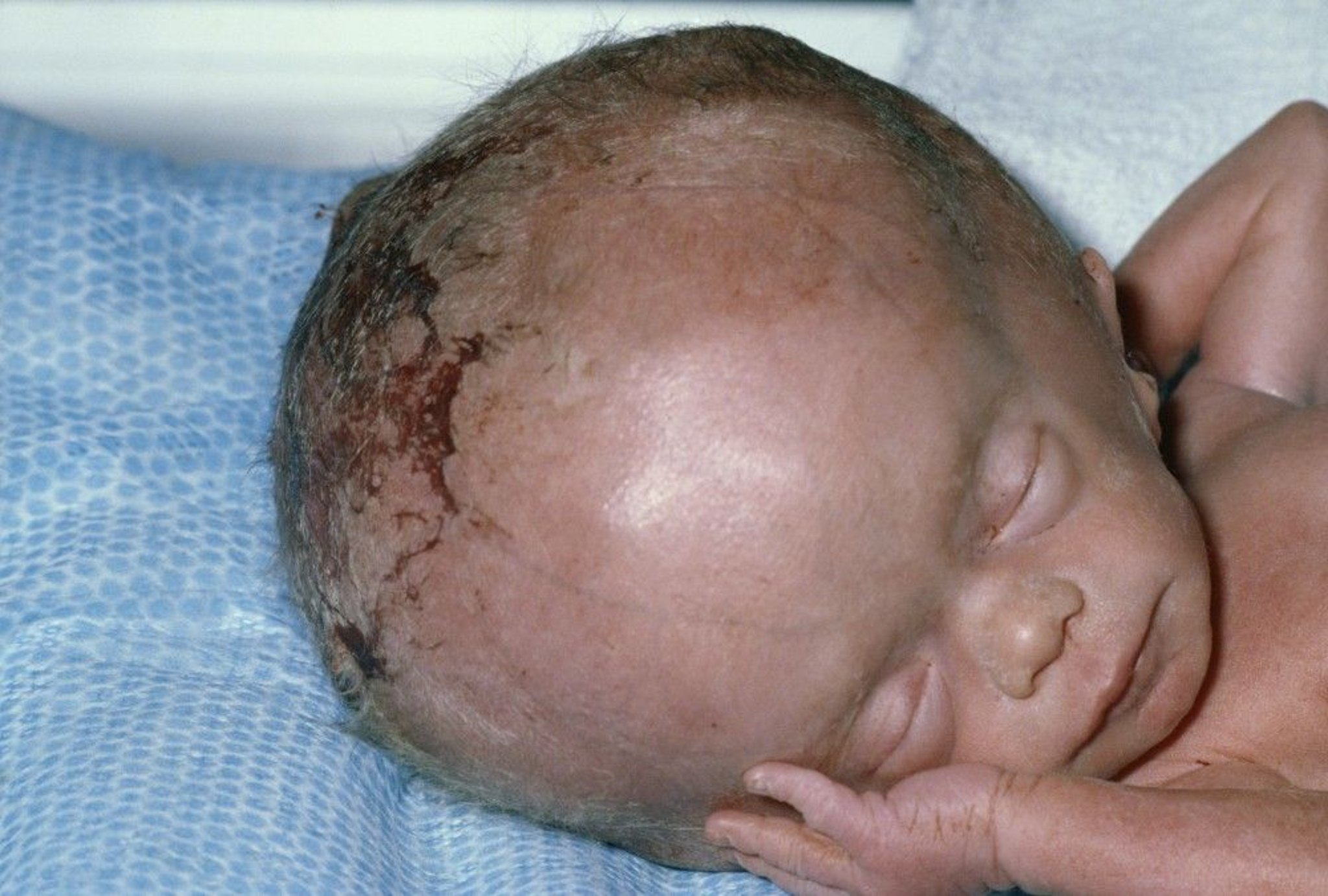
Hydrocephalus is the most common cause of abnormally large heads in neonates. Hydrocephalus that develops only after the fontanelles have closed does not increase head circumference or cause the fontanelles to bulge but can markedly and rapidly increase intracranial pressure.
Etiology of Hydrocephalus
Hydrocephalus can be either congenital or acquired from events during or after birth. It can be divided into two categories:
Obstructive hydrocephalus: Results from obstruction of cerebrospinal fluid (CSF) flow
Communicating hydrocephalus: Results from impaired resorption of CSF
The number of genes associated with congenital hydrocephalus continues to increase with new developments in clinical genetics. Mutations of many of these genes may cause hydrocephalus in conjunction with other neurodevelopmental abnormalities. Mutations in gene SLC12A6, which encodes a potassium chloride ion co-transporter, and in gene SLC12A7, a partner molecule, often also cause schizencephaly with hydrocephalus resulting from aqueductal stenosis, and manifest autosomal dominant inheritance. Genes LICAM1 and AP1S2 are X-linked, and genes CCDC88C and MPDZ are autosomal recessive (1).
Etiology reference
1. Shaheen R, Sebai MA, Patel N, et al: The genetic landscape of familial hydrocephalus. Ann Neurol 81(6):890–897, 2017. doi: 10.1002/ana.24964
Obstructive hydrocephalus
Obstruction most often occurs in the aqueduct of Sylvius but sometimes at the outlets of the 4th ventricle (Luschka and Magendie foramina).
The most common causes of obstructive hydrocephalus are
Aqueductal stenosis
Dandy-Walker malformation
Chiari II type malformation
Aqueductal stenosis is narrowing of the outflow pathway for CSF from the 3rd ventricle to the 4th ventricle. It may be either primary, or secondary to scarring or narrowing of the aqueduct resulting from a tumor, hemorrhage, or infection. Primary aqueductal stenosis may involve true stenosis (forking of the aqueduct into smaller, poorly functioning channels) or presence of a septum in the aqueduct. Primary aqueductal stenosis may be inheritable; there are many genetic syndromes, some of which are X-linked (thus male infants inherit the condition from otherwise unaffected mothers).
Dandy-Walker malformation comprises progressive cystic enlargement of the 4th ventricle in fetal life, resulting in complete or partial agenesis of the cerebellar vermis and hydrocephalus. It is likely related to a failure of formation of the foramen of Magendie in the fetus. Associated anomalies of the central nervous system are common, including agenesis of the corpus callosum and heterotopias. Dandy-Walker malformation accounts for 5 to 10% of cases of congenital hydrocephalus.
In Chiari II (formerly Arnold-Chiari), hydrocephalus occurs with spina bifida and syringomyelia. Significant elongation of the cerebellar tonsils in Chiari I type malformation or midline vermis in Chiari II causes them to protrude through the foramen magnum, with beaking of the colliculi and thickening of the upper cervical spinal cord.
Communicating hydrocephalus
Impaired resorption in the subarachnoid spaces usually results from meningeal inflammation, secondary either to infection or to blood in the subarachnoid space resulting from either subarachnoid hemorrhages or intraventricular hemorrhages, which are complications of delivery, particularly in preterm infants.
Symptoms and Signs of Hydrocephalus
Neurologic findings depend on whether intracranial pressure is increased.
In infants, symptoms of increased intracranial pressure include irritability, high-pitched cry, vomiting, lethargy, strabismus, and bulging fontanelle.
Older, verbal children may complain of headache, decreased vision, or both.
Papilledema is a late sign of increased intracranial pressure; its initial absence does not exclude hydrocephalus.
Consequences of chronic hydrocephalus may include precocious puberty in girls, learning disorders (eg, difficulties with attention, information processing, and memory), epilepsy, loss of vision, and impaired executive function (eg, problems with conceptualizing, abstracting, generalizing, reasoning, and organizing and planning information for problem-solving).
Diagnosis of Hydrocephalus
Prenatal ultrasonography
For neonates, cranial ultrasonography
For older infants and children, CT or MRI
Diagnosis of hydrocephalus is often made by routine prenatal ultrasonography.
After birth, diagnosis is suspected if routine examination reveals an increased head circumference; infants may have a bulging fontanelle or widely separated cranial sutures. Similar findings can result from intracranial space-occupying lesions (eg, subdural hematomas, porencephalic cysts, tumors). Macrocephaly may result from an underlying brain problem (eg, Alexander disease or Canavan disease), or it may be a benign, sometimes inherited, feature characterized by an increased amount of CSF surrounding a normal brain. Children suspected of having hydrocephalus require cranial imaging by CT, MRI, or ultrasonography (if the anterior fontanelle is open).
Cranial CT or ultrasonography is used to monitor progression of hydrocephalus once an anatomic diagnosis has been made. If seizures occur, electroencephalography may be helpful.
Treatment of Hydrocephalus
Sometimes observation or serial lumbar punctures
For severe cases, a ventricular shunt procedure
Treatment of hydrocephalus depends on etiology, severity, and whether hydrocephalus is progressive (ie, size of the ventricles increases over time relative to the size of the brain).
Mild, nonprogressive cases may be observed with serial imaging studies and measurement of head size. To temporarily reduce CSF pressure in infants, ventricular taps or serial lumbar punctures (if the hydrocephalus is communicating) may be used. These procedures are often used to treat and may reverse hydrocephalus with intracranial hemorrhage.
Progressive hydrocephalus usually requires a ventricular shunt. Shunts typically connect the right lateral ventricle to the peritoneal cavity or, rarely, to the right atrium via a plastic tube with a one-way, pressure-relief valve. When a shunt is first placed in an infant or older child whose fontanelles are closed, rapid withdrawal of fluid can cause subdural bleeding as the brain shrinks away from the skull. When the fontanelles are open, the skull can decrease in circumference to match the decrease in brain size; thus, some clinicians recommend an early decision regarding shunt placement so that it can be done before fontanelle closure.
In a third ventriculostomy, an opening is created endoscopically between the 3rd ventricle and the subarachnoid space, allowing CSF to drain. This procedure is often combined with ablation of the choroid plexus and is becoming more commonly used in the United States. It is particularly useful in countries where access to consistent neurosurgical care and follow-up is limited. In certain cases (eg, hydrocephalus caused by primary aqueductal stenosis), third ventriculostomy may be adequate primary treatment.
A ventricular shunt that goes to the subgaleal space may be used in infants as a temporary measure for those who may not require a more permanent shunt.
Although some children do not need the shunt as they age, shunts are rarely removed because of the risk of bleeding and trauma.
Fetal surgery to treat congenital hydrocephalus has not been successful.
Shunt complications
The type of ventricular shunt used depends on the neurosurgeon’s experience, but ventriculoperitoneal shunts cause fewer complications than ventriculoatrial shunts. Shunt complications include
Infection
Malfunction
Any shunt has a risk of infection. Manifestations include chronic fever, lethargy, irritability, headache, seizures, or other symptoms and signs of increased intracranial pressure; sometimes redness becomes apparent over the shunt tubing. Antibiotics effective against the organism infecting the shunt, which may include skin flora, are given, and typically the shunt must be removed and replaced.
Shunts can malfunction because of a mechanical obstruction (typically blockage at the ventricular end) or because of fracture of the tubing. In either case, intracranial pressure can increase, which, if sudden, can be a medical emergency. Children most often present with headache, vomiting, lethargy, irritability, esotropia, or paralysis of upward gaze. Seizures may occur. If the obstruction is gradual, more subtle symptoms and signs can occur, such as irritability, poor school performance, and lethargy, which may be mistaken for depression.
To assess shunt function, a shunt series (x-rays of the shunt tubing) and neuroimaging studies are done. The ability to compress the bulb that is present on many shunt systems is not a reliable sign of shunt function.
After the shunt is placed, head circumference and development are assessed, and imaging is done periodically.
Key Points
Hydrocephalus is usually caused by obstruction to the normal flow of cerebrospinal fluid (CSF) but can be due to impaired resorption of CSF.
If the disorder occurs before the cranial sutures have fused, the head may be enlarged, with bulging fontanelles.
Neurologic symptoms develop mainly if intracranial pressure increases; infants may have irritability, high-pitched cry, vomiting, lethargy, and strabismus.
Diagnose using ultrasonography prenatally and in neonates; use MRI or CT for older children.
Treat with observation or serial lumbar punctures or a ventricular shunt procedure depending on the etiology and severity and progression of symptoms.


