

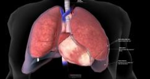
Pleural effusion is the abnormal accumulation of fluid in the pleural space (the area between the 2 layers of the thin membrane that covers the lungs).
Fluid can accumulate in the pleural space as a result of use of some medications or because of a large number of disorders, including infections; tumors; injuries; heart, kidney, or liver failure; and blood clots in the lung blood vessels (pulmonary emboli).
Symptoms may include difficulty breathing and chest pain particularly when breathing and coughing.
Diagnosis is by chest x-rays or ultrasound examination, laboratory testing of the fluid, and often computed tomography angiography.
Large amounts of fluid are drained with a tube inserted into the chest.
(See also Overview of Pleural and Mediastinal Disorders.)
Normally, only a thin layer of fluid separates the 2 layers of the pleura. An excessive amount of fluid may accumulate for many reasons, including heart failure, cirrhosis, pneumonia, and cancer.
A large number of disorders may cause pleural effusion. Some of the more common causes (listed as roughly the most common to least common) include the following:
Tumors
Surgery, such as recent coronary artery bypass surgery
Injury to the chest
Systemic lupus erythematosus (lupus)
Nephrotic syndrome (protein in the urine and high blood pressure)
Medications such as hydralazine, procainamide, isoniazid, phenytoin, chlorpromazine, methysergide, interleukin-2, nitrofurantoin, bromocriptine, dantrolene, and procarbazineMedications such as hydralazine, procainamide, isoniazid, phenytoin, chlorpromazine, methysergide, interleukin-2, nitrofurantoin, bromocriptine, dantrolene, and procarbazine
Types of fluid
Depending on the cause, the fluid may be either:
Rich in protein (exudate)
Watery (transudate)
Doctors use this distinction to help determine the cause. For example, heart failure and cirrhosis are common causes of watery fluid in the pleural space. Pneumonia, cancer, and viral infections are common causes of pleural effusion with an exudative fluid.
Blood in the pleural space (hemothorax) usually results from a chest injury. Rarely, a blood vessel ruptures into the pleural space when no injury has occurred, or a bulging area in the aorta (aortic aneurysm) leaks blood into the pleural space.
Pus in the pleural space (empyema) can accumulate when pneumonia or a lung abscess spreads into the space. Empyema may also complicate an infection due to chest wounds, chest surgery, rupture of the esophagus, or an abscess in the abdomen.
Lymphatic (milky) fluid in the pleural space (chylothorax) is caused by an injury to the main lymphatic duct in the chest (thoracic duct) or by a blockage of the duct by a tumor.
Symptoms of Pleural Effusion
Many people with pleural effusion have no symptoms at all. The most common symptoms, regardless of the type of fluid in the pleural space or its cause, are:
Shortness of breath
Chest pain
Chest pain is usually of a type called pleuritic pain (the term pleurisy is no longer or only rarely used). Pleuritic pain may be felt only when the person breathes deeply or coughs, or it may be felt continuously but may be worsened by deep breathing and coughing. The pain is usually felt in the chest wall right over the site of the inflammation or infection that caused the effusion. However, the pain may be felt also or only in the upper abdominal region or neck and shoulder, which is called referred pain (see figure ). Pleuritic pain can be caused by disorders other than pleural effusion, including pneumonia, rib fractures, blood clots in the lung, viral infections, pancreatitis, rheumatoid arthritis, and lupus.
Pleuritic chest pain due to a pleural effusion may disappear as fluid accumulates. Large amounts of fluid can cause difficulty in expanding one or both lungs when breathing, causing shortness of breath.
Diagnosis of Pleural Effusion
Chest x-ray, ultrasound examination, or both
Laboratory tests done on a sample of the fluid
Sometimes computed tomography (CT)
A chest x-ray, which shows fluid in the pleural space, is usually the first step in making the diagnosis. However, small amounts of fluid may not be visible on a chest x-ray.
An ultrasound of the chest can also be done to help doctors identify small accumulations of fluid.
Doctors may do thoracentesis. In this procedure, doctors use a needle to remove a specimen of the fluid for examination. The appearance of the fluid may help doctors determine its cause. Certain laboratory tests evaluate the chemical composition of the fluid and determine the presence of bacteria, including the bacteria that cause tuberculosis. The fluid specimen is also examined for the number and types of cells and for the presence of cancerous cells.
If these tests cannot identify the cause of the pleural effusion, other tests may be done.
CT scan more clearly shows the lung and the fluid and may show evidence of pneumonia, a mediastinal mass, a lung abscess, or a tumor that could be causing fluid to accumulate. Sometimes a radiopaque dye is injected during CT (CT angiography or venography) to look for problems with the pleura or with blood vessels, including pulmonary embolism.
If a serious diagnosis still seems possible, doctors may insert a viewing tube into the chest (called thoracoscopy). Occasionally, doctors need to obtain a sample (biopsy) of the pleura and/or lung. In some people with pleural effusion, the cause is not obvious after initial testing, and in some people a cause is never found, even after extensive testing.
Treatment of Pleural Effusion
Treatment of the disorder causing pleural effusion
Drainage of large pleural effusions
Small pleural effusions that are not causing symptoms may not require treatment, although the underlying disorder must be treated. Sometimes the person is given analgesics until the fluid is drained or drains away on its own.
Larger pleural effusions, especially those that cause shortness of breath, may require drainage. Usually, drainage dramatically relieves shortness of breath. Often, fluid can be drained using thoracentesis. An area of skin between 2 lower ribs is anesthetized, then a small needle is inserted and gently pushed deeper until it reaches the fluid. A catheter (thin flexible tube) is often guided over the needle into the fluid to lessen the chance of puncturing the lung and causing a collapsed lung (pneumothorax), or damaging other organs or structures. Although thoracentesis is usually done for diagnostic purposes, doctors may use this procedure to remove enough fluid at one time to relieve the person's shortness of breath.
When large amounts of fluid must be removed, a tube (chest tube) may be inserted through the chest wall. After numbing the area by injecting a local anesthetic, doctors insert a thin flexible tube into the chest between 2 ribs. Then doctors connect the tube to a water-sealed drainage system that prevents air from leaking into the pleural space. A chest x-ray is taken to check the tube’s position. Drainage can be blocked if the chest tube is incorrectly positioned or becomes kinked. If the fluid is very thick or full of clots, it may not flow out.
Effusions caused by pneumonia
When an accumulation of fluid is due to pneumonia, antibiotics are needed. Doctors also typically remove a sample of the fluid for examination and testing. If the fluid is pus or has certain other characteristics, the fluid needs to be drained, usually with a chest tube. If the fluid is partitioned into separate compartments by scars that have developed within the pleural space, drainage is more difficult. Sometimes fibrinolytic medications, which dissolve blood clots, plus a medication that can help thin effusions that are thick (dornase alfa) are instilled into the pleural space to help drainage, which may avoid the need for surgery. (To be effective, both fibrinolytic medications and When an accumulation of fluid is due to pneumonia, antibiotics are needed. Doctors also typically remove a sample of the fluid for examination and testing. If the fluid is pus or has certain other characteristics, the fluid needs to be drained, usually with a chest tube. If the fluid is partitioned into separate compartments by scars that have developed within the pleural space, drainage is more difficult. Sometimes fibrinolytic medications, which dissolve blood clots, plus a medication that can help thin effusions that are thick (dornase alfa) are instilled into the pleural space to help drainage, which may avoid the need for surgery. (To be effective, both fibrinolytic medications anddornase alfa must be used.)
If surgery is needed, it can be done by using a procedure called video-assisted thoracoscopic debridement or by making an incision through the chest wall (thoracotomy). During surgery, any thick peels of fibrous material over the lung surface are removed to allow the lung to expand normally.
Effusions caused by cancers
Fluid accumulation caused by cancers of the pleura may be difficult to treat because fluid often reaccumulates rapidly. Draining the fluid and giving antitumor medications sometimes prevents further fluid accumulation. A small tube can be left in the chest so that the fluid can be drained periodically into vacuum bottles. But if fluid continues to accumulate, sealing the pleural space (pleurodesis) may be helpful. For pleurodesis, all fluid is drained through a tube, which is then used to administer a pleural irritant, such as a doxycycline solution, bleomycin, or a talc mixture, into the space. The irritant seals the two layers of pleura together, so that no room remains for additional fluid to accumulate. Pleurodesis can also be done using thoracoscopy.Fluid accumulation caused by cancers of the pleura may be difficult to treat because fluid often reaccumulates rapidly. Draining the fluid and giving antitumor medications sometimes prevents further fluid accumulation. A small tube can be left in the chest so that the fluid can be drained periodically into vacuum bottles. But if fluid continues to accumulate, sealing the pleural space (pleurodesis) may be helpful. For pleurodesis, all fluid is drained through a tube, which is then used to administer a pleural irritant, such as a doxycycline solution, bleomycin, or a talc mixture, into the space. The irritant seals the two layers of pleura together, so that no room remains for additional fluid to accumulate. Pleurodesis can also be done using thoracoscopy.
Chylothorax
Treatment of chylothorax focuses on eliminating the leakage from the lymphatic duct. Such treatment may consist of surgery, chemotherapy, or radiation therapy for a tumor that is blocking lymph flow.


