

An artery in the retina (the transparent, light-sensitive structure at the back of the eye) may become blocked, causing sudden, painless loss of vision.
Doctors typically make the diagnosis by looking in the eye with an ophthalmoscope and sometimes by testing.
Treatments are usually unsuccessful in restoring vision.
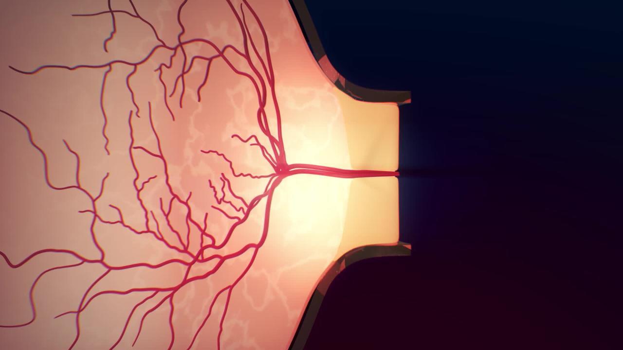
The central retinal artery is the main vessel that supplies blood to the retina. This artery can become completely blocked by an embolism or thrombosis (formation of a blood clot in the artery). Blockage may occur in the main artery or in its branches. (See also Overview of Retinal Disorders.)

An embolism is a collection of solid material that floats in the bloodstream until it gets stuck in and blocks a blood vessel. The material that forms an embolus can come from a piece of atherosclerotic plaque from an artery in the neck, fat, infected material from an infected heart valve (endocarditis), or a noncancerous (benign) tumor in a heart chamber (atrial myxoma).
Giant cell arteritis, an inflammation of the blood vessels, is also a possible cause of retinal artery blockage, generally in people older than 50 years, particularly those who have polymyalgia rheumatica.
Sometimes the cause of the blockage is unknown.
Symptoms of Retinal Artery Blockage
The affected eye has a sudden and severe but painless loss of vision over the entire field of vision. Sometimes only a part of the field of vision is affected.
Blockage of the central retinal artery may also cause growth of abnormal blood vessels on the retina or iris. Sometimes these abnormal blood vessels bleed or cause a painful type of glaucoma (called neovascular glaucoma). In neovascular glaucoma, abnormal blood vessels that have formed in the iris close the space between the iris and the cornea, blocking the drainage of fluid from the eye and causing buildup of pressure in the eye.
Diagnosis of Retinal Artery Blockage
A doctor's examination of the eye
Fluorescein angiographyFluorescein angiography
Optical coherence tomography
Sometimes echocardiography, Doppler ultrasonography, and/or blood tests
Using an ophthalmoscope, doctors can see changes in blood vessels and the retina. If the central retinal artery is blocked, the retina may appear pale.
Fluorescein angiography helps determine the extent of damage to the retina and helps the doctor plan treatment. In this procedure, a doctor injects dye into a vein in the arm and then photographs the retina. Optical coherence tomography (an imaging study) can help show that the retina is swollen, which is common.
Once retinal artery blockage has been diagnosed, doctors need to search for a source of an embolus. They do tests such as echocardiography and carotid Doppler ultrasonography. They may also do blood tests to diagnose giant cell arteritis.
If people experience sudden brief loss of vision in one eye and doctors think this is due to a clot that is blocking the retinal artery, treatment is started immediately rather than delayed for imaging studies.
Treatment of Retinal Artery Blockage
Prevention by controlling risk factors
Sometimes measures to lower pressure in the eye, including eyedrops, eye massage, or removing fluid from the eye with a needle
Sometimes laser treatment of abnormal or bleeding blood vessels
For giant cell arteritis, corticosteroids
Because treatments tend not to be effective, preventing retinal artery blockages by controlling risk factors (for example, high blood pressure, diabetes, and other risk factors for atherosclerosis) is desirable.
When blockage of a central retinal artery does occur, immediate treatment is often given in an attempt to unblock the retinal artery. However, treatments are rarely effective. Pressure inside the eye sometimes can be lowered by giving medications that lower blood pressure (such as timolol eye drops or acetazolamide taken by mouth).When blockage of a central retinal artery does occur, immediate treatment is often given in an attempt to unblock the retinal artery. However, treatments are rarely effective. Pressure inside the eye sometimes can be lowered by giving medications that lower blood pressure (such as timolol eye drops or acetazolamide taken by mouth).
Pressure inside the eye may be lowered by massage or by a procedure called anterior chamber paracentesis (which removes fluid from the eye with a needle). Lowering the pressure inside the eye may dislodge a blood clot or embolus and allow it to enter a smaller branch of the vessel, thereby reducing the area of damage to the retina.
People with suspected giant cell arteritis are given high-dose corticosteroids either by mouth or by vein as soon as possible.
Laser treatment may be used to destroy abnormal blood vessels to treat or prevent neovascular glaucoma, prevent further vision loss from bleeding within the eye, or both. However, treatment of neovascular glaucoma is difficult.
Because people with central retinal artery blockage have an increased risk of stroke, doctors immediately refer them to a specialized stroke center for further evaluation.
Prognosis for Retinal Artery Blockage
If the blockage occurred in a branch of the central retinal artery, people may maintain good to fair vision.
If the blockage occurred in the central retinal artery itself, vision loss is often profound, even with treatment.
Once the retinal tissue becomes permanently damaged, which can happen as quickly as 90 minutes after the blockage, vision loss is usually permanent.
If giant cell arteritis is the cause of the retinal artery blockage, prompt diagnosis and treatment may allow people to regain some lost vision and be protected from damage to the other eye.
People with retinal artery blockage may have other blockages affecting arteries that supply the brain. These blockages increase the risk of stroke, particularly in the weeks following a central retinal artery blockage.
More Information
The following English-language resource may be useful. Please note that THE MANUAL is not responsible for the content of this resource.
National Eye Institute: A resource for learning about eye health (in English and Spanish) for adults and children, as well as for providing access to outreach campaigns.


