


MRI provides better resolution of neural structures than CT. This difference is most significant clinically for visualizing the following:
Cranial nerves
Brain stem lesions
Abnormalities of the posterior fossa
Spinal cord
CT images of these regions are often marred by bony streak artifacts. MRI is especially valuable for identifying spinal abnormalities (eg, tumor, abscess) compressing the spinal cord and requiring emergency intervention. Also, MRI is better for detecting demyelinating plaques, early infarction, subclinical brain edema, cerebral contusions, incipient transtentorial herniation, abnormalities of the craniocervical junction, and syringomyelia.

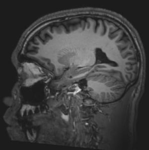
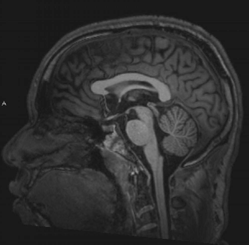
© 2017 Elliot K. Fishman, MD.
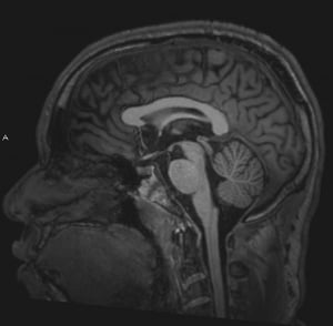
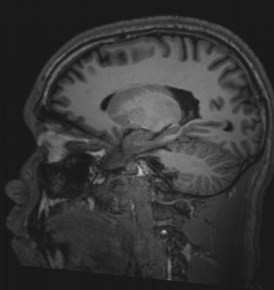
© 2017 Elliot K. Fishman, MD.

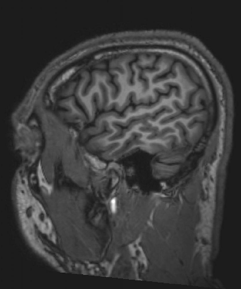
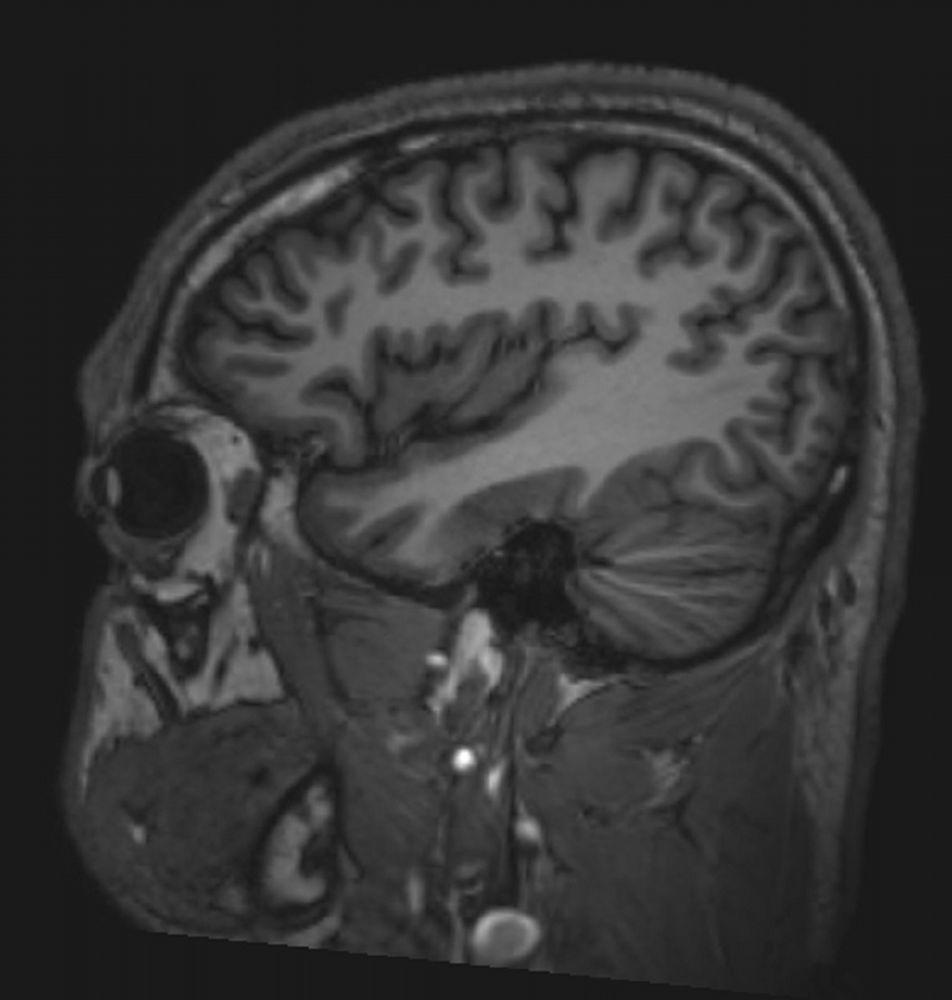
© 2017 Elliot K. Fishman, MD.

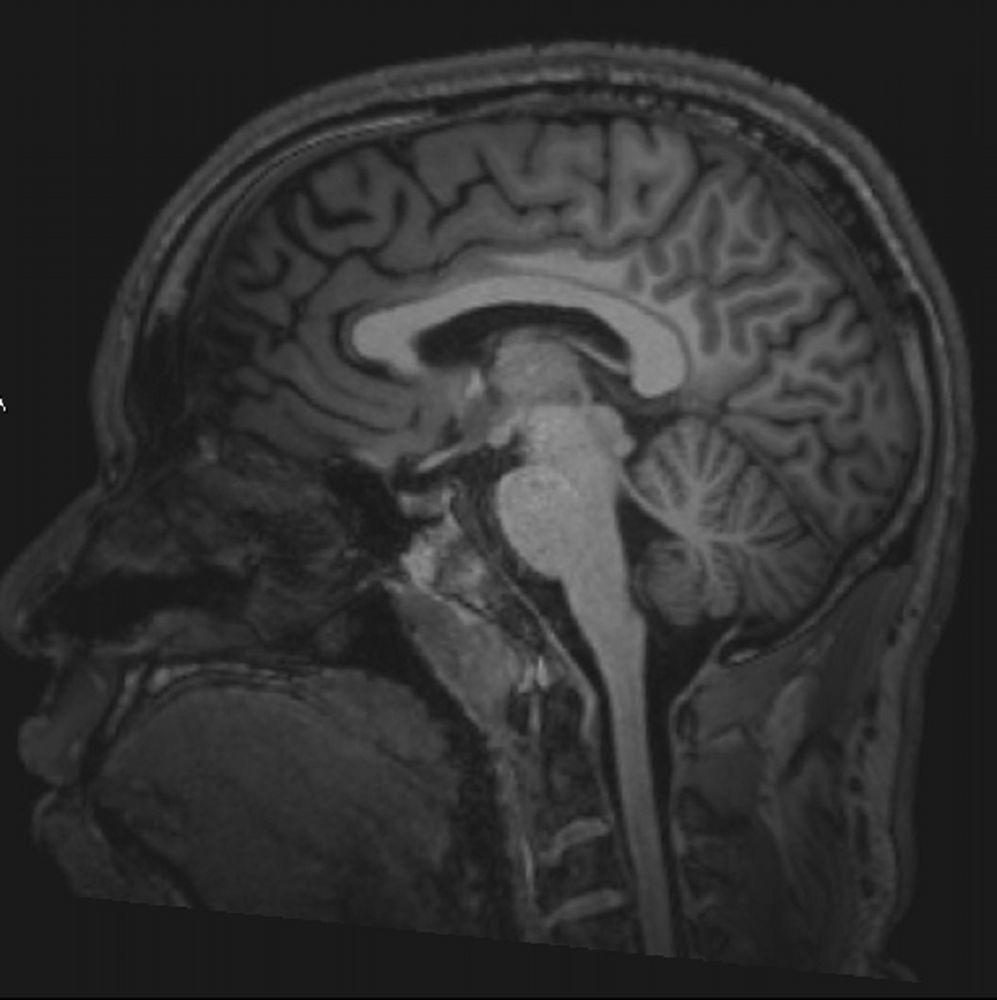
© 2017 Elliot K. Fishman, MD.

© 2017 Elliot K. Fishman, MD.

© 2017 Elliot K. Fishman, MD.


© 2017 Elliot K. Fishman, MD.

© 2017 Elliot K. Fishman, MD.


© 2017 Elliot K. Fishman, MD.

© 2017 Elliot K. Fishman, MD.
MRI is contraindicated if patients
Have had a pacemaker or cardiac or carotid stents for < 6 weeks
Have ferromagnetic aneurysm clips or other metallic objects that may overheat or be displaced within the body by the intense magnetic field
Visualization of inflammatory, demyelinated, and neoplastic lesions may require enhancement with IV paramagnetic contrast agents (eg, gadolinium). Although gadolinium is thought to be much safer than contrast agents used with CT, nephrogenic systemic fibrosis (nephrogenic fibrosing dermopathy) has been reported in patients with impaired renal function and acidosis. Before using gadolinium in patients with renal disease, clinicians should consult with a radiologist and a nephrologist.
There are several MRI techniques; choice of technique depends on the specific tissue, location, and suspected disorder:
Diffusion-weighted imaging (DWI) allows rapid, early detection of ischemic stroke and helps distinguish cerebral abscess from tumor. It can also help diagnose Creutzfeld-Jacob disease.
Perfusion-weighted imaging (PWI) can detect areas of hypoperfusion in early ischemic stroke but cannot yet reliably distinguish areas with benign oligemia from those with injurious hypoperfusion due to infarction.
Diffusion tensor imaging (DTI) is an extension of DWI that can show white matter tracts in 3 dimensions (tractography) and can be used to monitor the integrity of CNS tracts affected by aging and disease.
Flow-attenuated inversion recovery (FLAIR) is used to distinguish demyelinating lesions, such as those seen in multiple sclerosis, from a water signal coming from CSF; with this technique, the CSF looks dark and the demyelinating lesion looks white.
Double inversion recovery (DIR), used in research centers, can detect demyelination of gray matter better than other MRI techniques; gray matter demyelination is now considered common in multiple sclerosis.
Functional MRI (fMRI) shows which brain regions are activated (shown by increased flow of oxygenated blood) by a specific cognitive or motor task, but its clinical use is still being defined.
Magnetic resonance angiography (MRA) uses MRI with or without a contrast agent to show cerebral vessels and major arteries and their branches in the head and neck. Although MRA has not replaced cerebral angiography, it is used when cerebral angiography cannot be done (eg, because the patient refuses or has increased risk). As a check for stroke, MRA tends to exaggerate severity of arterial narrowing and thus does not usually miss occlusive disease of large arteries. It provides better images than CT angiography when cerebral vessel dissection is suspected.
Susceptibility-weighted angiography (SWAN) can be useful in evaluating bleeding. It provides better visualization of both large and small blood vessels, microhemorrhages, and deposits of calcium and iron in the brain. It can also show tiny blood vessels (eg, venules) that are most often seen in the center of demyelinating lesions in patients with multiple sclerosis and thus distinguishes lesions due to multiple sclerosis from ischemic lesions.
Magnetic resonance venography (MRV) uses MRI to show the major veins and dural sinuses of the cranium. MRV obviates the need for cerebral angiography in diagnosing cerebral venous thrombosis and is useful for monitoring thrombus resolution and guiding the duration of anticoagulation.
Magnetic resonance spectroscopy can measure metabolites in the brain regionally to distinguish tumors from abscess or stroke.


