


The main purpose of the respiratory system is to take in oxygen and eliminate carbon dioxide through the lungs. Oxygen, which is in the air, is breathed into the lungs (inhaled). Oxygen is necessary for the body to produce energy and sustain life. Carbon dioxide, a waste product of energy production, is dangerous if it builds up, and must be removed from the body by the lungs, back into air that is breathed out (exhaled). The respiratory system works closely with the circulatory system, which brings the oxygen from the lungs to the body's organs and removes carbon dioxide from the organs and takes it to the lungs.
The respiratory system also helps with maintaining body temperature (by regulating the temperature of inhaled air), eliminating water from the body (by water vapor in exhaled air), removing dust and microorganisms from incoming air, clearing mucous or other substances from the lungs (by coughing and the movement of tiny hairlike structures called cilia), facilitating the sense of smell (by passing air over the organs of smell in the nose), and producing sound (in the voice box, or larynx).
The respiratory system starts at the nose and mouth and continues through the airways and the lungs. Air enters the respiratory system through the nose and mouth and passes down the throat (pharynx) and through the larynx. The entrance to the larynx is covered by a small flap of tissue, the epiglottis, that automatically closes during swallowing, thus preventing food or drink from entering the airways.
The trachea (windpipe) is the largest airway. The trachea branches into 2 smaller airways: the left and right mainstem [or main] bronchi.
Each lung is divided into sections (lobes): 3 in the right lung and 2 in the left lung. The left lung is a little smaller than the right lung because it shares space in the left side of the chest with the heart.
Inside the Lungs and Airways

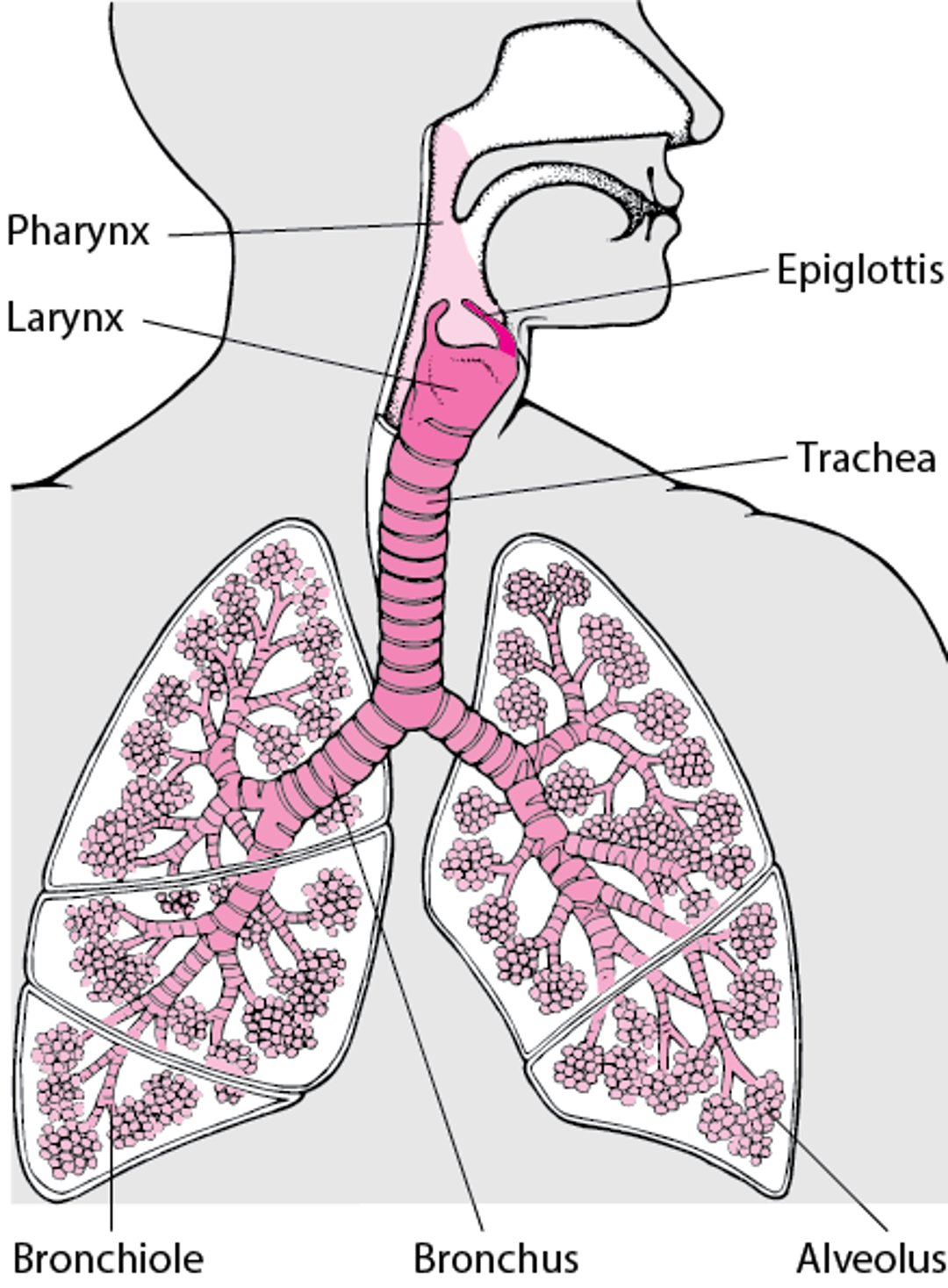
The bronchi themselves branch many times into smaller airways, ending in the narrowest airways (bronchioles), which are as small as one-half of a millimeter (or 2/100 of an inch) across. The airways resemble an upside-down tree, which is why this part of the respiratory system is often called the bronchial tree. Large airways are held open by semiflexible, fibrous connective tissue called cartilage. Smaller airways are supported by the lung tissue that surrounds and is attached to them. The walls of the smaller airways have a thin, circular layer of smooth muscle. The airway muscle can relax or contract, thus changing airway size.
Thousands of alveoli (small air sacs) are at the end of each bronchiole. Although each individual alveolus is less than half a millimeter (less than 0.02 inches) in diameter, together, the millions of alveoli of the lungs form a surface of more than 100 square meters (1111 square feet). Within the alveolar walls is a dense network of tiny blood vessels called capillaries. The extremely thin barrier between air and capillaries allows oxygen to move from the alveoli into the blood and allows carbon dioxide to move from the blood in the capillaries into the air in the alveoli.
The pleura is a slippery membrane that covers the lungs as well as the inside of the chest wall. It allows the lungs to move smoothly during breathing and as the person moves. Normally, the 2 layers of the pleura have only a small amount of lubricating fluid between them. The 2 layers glide smoothly over each other as the lungs change size and shape.


