

In tetralogy of Fallot, four specific heart defects occur together.
This condition includes four heart defects that can lead to oxygen-poor blood going directly to the body.
Symptoms include mild to severe cyanosis (a bluish discoloration of the skin due to lack of oxygen in the blood), life-threatening attacks of intense cyanosis due to a rapid drop in oxygen in the blood, and a heart murmur (a sound created by turbulent blood flow through narrowed or leaking heart valves or through abnormal heart structures).
The diagnosis is suspected based on a characteristic murmur and on the presence of cyanosis and is confirmed based on the results of echocardiography.
Surgery is required to correct the defect.
(See also Overview of Heart Defects.)
The four heart defects are
A narrowing of the outflow passage from the right side of the heart
A large ventricular septal defect (a hole in the wall that separates the left from the right ventricle)
Displacement of the aorta that allows oxygen-poor blood to flow directly from the right ventricle to the aorta (causing a right-to-left shunt), which is also called overriding aorta
A thickening of the wall of the right ventricle
Tetralogy of Fallot: Four Defects

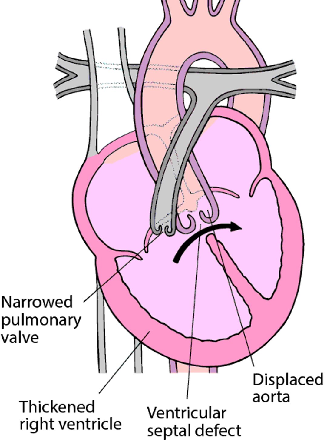
In infants with tetralogy of Fallot, the narrowed passage from the right ventricle restricts blood flow to the lungs. The restricted blood flow causes the oxygen-poor blood in the right ventricle to pass through the septal defect to the left ventricle and into the aorta (right-to-left shunt). The more oxygen-poor blood (which is blue) that flows to the body, the bluer the body appears.
Infants with severe or complete blockage of blood flow from the right side of the heart may depend on having an open ductus arteriosus for survival. The ductus arteriosus is a blood vessel in the fetus that connects the two great arteries leaving the heart, the pulmonary artery and the aorta (see Normal Fetal Circulation). After birth, the ductus arteriosus is no longer needed and usually closes within the first days of life. However, if the ductus stays open after birth in infants with severe tetralogy, some blood from the aorta can flow back into the lungs through the open ductus and thus pick up oxygen.
Symptoms of Tetralogy of Fallot
The main symptom is
Cyanosis (a bluish discoloration of the skin), which can be mild or severe
Infants with tetralogy of Fallot usually have a heart murmur. A heart murmur is a sound created by turbulent blood through narrowed or leaking heart valves or through abnormal heart structures.
Some children have life-threatening attacks (hypercyanosis or "tet" spells) during which cyanosis suddenly worsens without obvious cause or in response to activity, such as crying or having a bowel movement. The child becomes very irritable and short of breath and may lose consciousness. The heart murmur often disappears during these spells.
Diagnosis of Tetralogy of Fallot
Echocardiography
Doctors suspect tetralogy of Fallot based on a characteristic harsh murmur that can be heard with a stethoscope. In addition, oxygen levels are usually lower than normal when checked with a skin sensor (pulse oximetry).
Echocardiography (ultrasonography of the heart) shows the four heart defects and confirms the diagnosis.
Electrocardiography (ECG) and chest x-rays are typically done. The ECG may be normal in the first month or two of life, but then will show increased thickness of the right ventricle. The chest x-ray usually shows an abnormally shaped heart.
Treatment of Tetralogy of Fallot
Sometimes a medication, such as a prostaglandin, to keep the ductus arteriosus open in the first week of life
For hypercyanotic spells, positioning, calming, oxygen, and sometimes medications and/or fluids given by vein,
An oral beta-blocker medication may be used after a hypercyanotic spell resolves until surgery can be done
Surgery
In infants who depend on an open ductus arteriosus for survival, giving a prostaglandin by vein to maintain an open ductus arteriosus can be lifesaving. Keeping the ductus arteriosus open sends extra blood to the lungs and increases the level of oxygen in the infant's blood.
Hypercyanotic spells
When an infant has a hypercyanotic spell, the infant may breathe more easily when the knees are close to the chest (knee-chest position). Interestingly, older children with tetralogy of Fallot will naturally do the same thing by squatting down, which helps to push more blood to the lungs and makes them feel better. Calming the infant and giving oxygen also help. If these measures do not work, morphine, fluids given by vein (intravenously), and medications such as a beta-blocker (for example, propranolol), or phenylephrine may be given to improve blood flow to the lungs. When an infant has a hypercyanotic spell, the infant may breathe more easily when the knees are close to the chest (knee-chest position). Interestingly, older children with tetralogy of Fallot will naturally do the same thing by squatting down, which helps to push more blood to the lungs and makes them feel better. Calming the infant and giving oxygen also help. If these measures do not work, morphine, fluids given by vein (intravenously), and medications such as a beta-blocker (for example, propranolol), or phenylephrine may be given to improve blood flow to the lungs.
Any infant or child experiencing hypercyanotic spells should have heart surgery promptly. A doctor may give the infant propranolol to decrease the risk of future spells if surgery cannot be done immediately.Any infant or child experiencing hypercyanotic spells should have heart surgery promptly. A doctor may give the infant propranolol to decrease the risk of future spells if surgery cannot be done immediately.
Surgery
In infants with tetralogy of Fallot, the defects need repair with surgery. If oxygen levels are low or infants have hypercyanotic spells, surgery is done in early infancy. If children have few symptoms, surgery is sometimes delayed until later in infancy.
If infants have low birth weight or have complex defects, doctors may use less invasive procedures to keep blood flowing to the lungs until surgery can be done. For example, they may use a synthetic blood vessel (a shunt) to connect the aorta to a lung artery. This procedure routes blood to the lungs so that more blood can obtain oxygen before it goes to the rest of the body. Another option can be done during cardiac catheterization, in which a thin tube (catheter) with an expandable flexible tube (stent) at its tip is passed through a blood vessel in the leg into the heart. The stent is expanded in the heart to enlarge the outflow to the lungs, which helps increase levels of oxygen in the blood.
During surgery, the ventricular septal defect is closed, the narrowed passageway from the right ventricle and the narrowed pulmonary valve are widened, and the patent ductus arteriosus is closed.
Children need to take antibiotics before visits to the dentist and before certain surgeries (such as on the respiratory tract) before and usually after surgical repair. These antibiotics are used to prevent a serious heart infection called endocarditis.
More Information
The following English-language resources may be useful. Please note that THE MANUAL is not responsible for the content of these resources.
American Heart Association: Common Heart Defects: Provides an overview of common birth defects of the heart for parents and caregivers
American Heart Association: Infective Endocarditis: Provides an overview of infective endocarditis, including summarizing antibiotic use, for parents and caregivers


