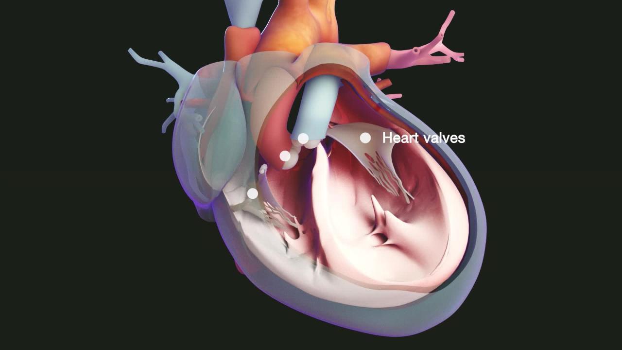
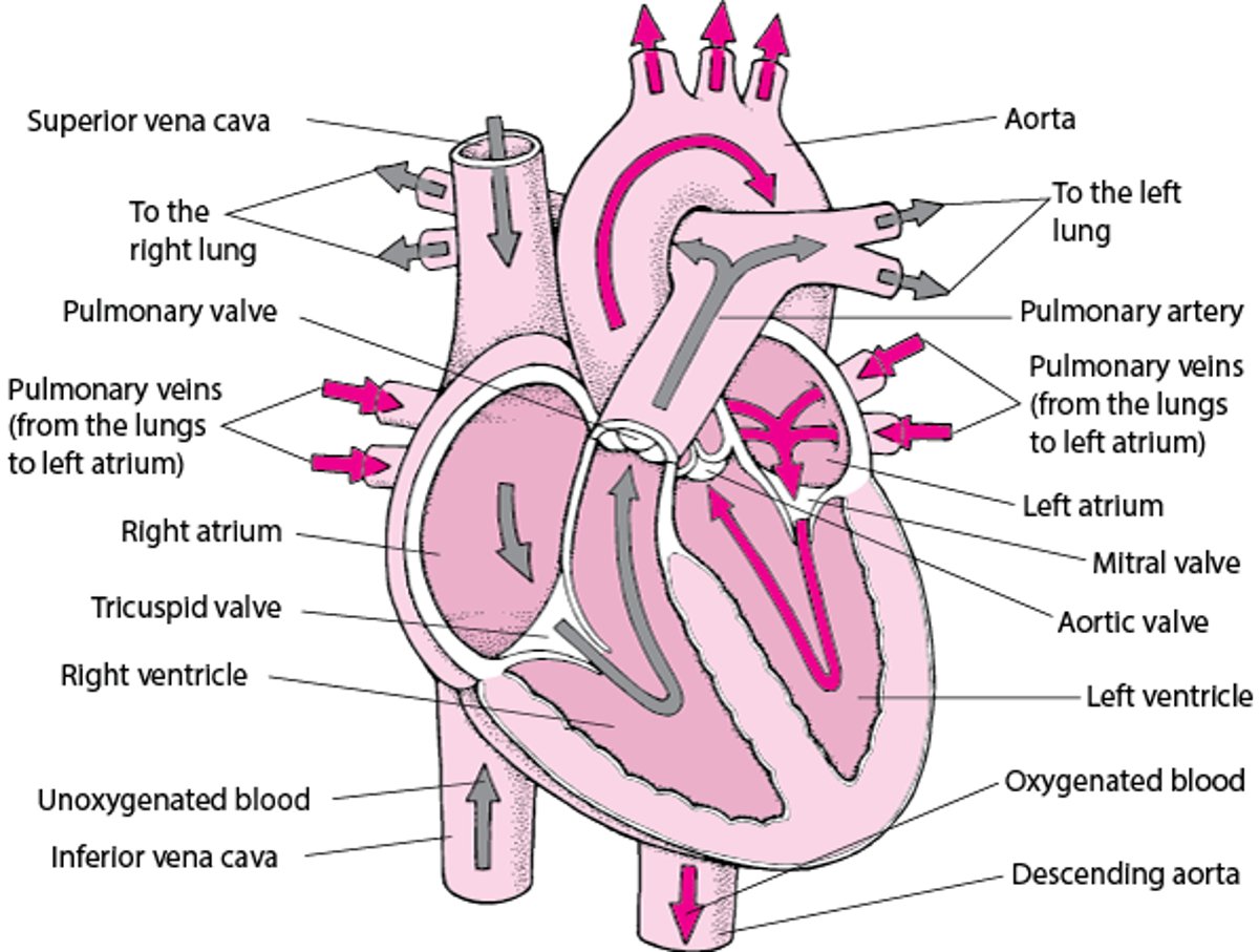
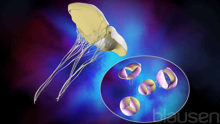
Heart valves regulate the flow of blood through the heart's four chambers—two small, round upper chambers (atria) and two larger, cone-shaped lower chambers (ventricles). Each ventricle has a one-way "in" (inlet) valve and a one-way "out" (outlet) valve. Each valve consists of flaps (cusps or leaflets) that open and close like one-way swinging doors.
In the right ventricle, the inlet valve is the tricuspid valve, which opens from the right atrium, and the outlet valve is the pulmonary (pulmonic) valve, which opens into the pulmonary artery.

In the left ventricle, the inlet valve is the mitral valve, which opens from the left atrium, and the outlet valve is the aortic valve, which opens into the aorta.
The mitral and tricuspid valves are held in place by tough, fibrous strings (chordae tendineae) that are connected to thin muscles (papillary muscles) that attach to the walls of the ventricles.
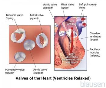
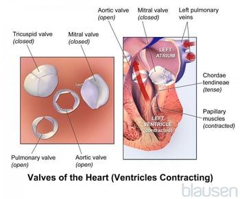
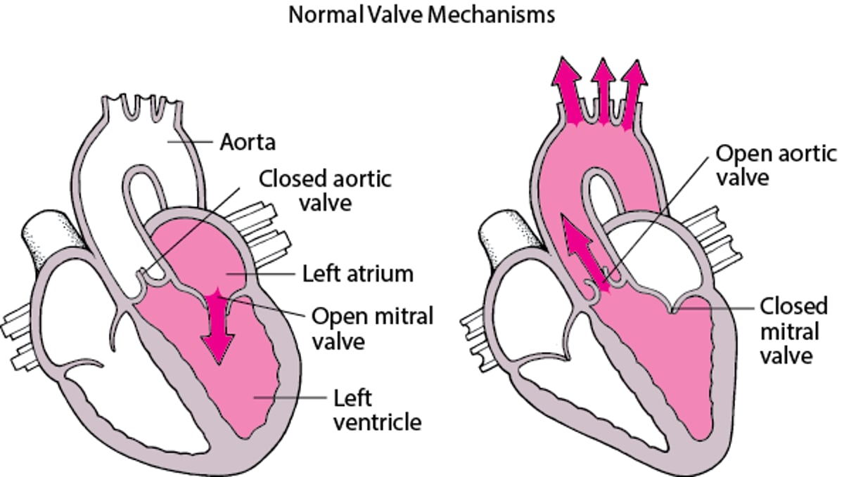
A Look Into the Heart
This cross-sectional view of the heart shows the direction of normal blood flow. |

Changes in heart valves with aging
As people age, the mitral and aortic valves thicken. The aorta becomes stiffer, which increases blood pressure and stress on the aortic valve, and the heart requires additional oxygen to pump blood effectively. These age-related changes may lead to symptoms and complications in older people with heart disease.
Heart valve malfunction
The heart valves can malfunction by
Leaking (termed regurgitation, incompetence, or insufficiency)
Not opening adequately and thus partially blocking the flow of blood through the valve (termed stenosis)
Either problem can greatly interfere with the heart's ability to pump blood. Sometimes a valve has both problems. Faulty valves generally create murmurs and other abnormal heart sounds that a doctor can hear with a stethoscope. Faulty valves can be identified by using echocardiography. Often, minor degrees of regurgitation are not detected with a stethoscope but are detected during echocardiography. Doctors often regard some murmurs they hear and some minor regurgitation they observe with echocardiography as this is a normal finding.
Most faulty valves are not severely abnormal, and they do not affect the person in any way. Symptoms may include leg swelling, shortness of breath, palpitations, fatigue, chest pain, and fainting.
Doctors often do periodic check-ups because a few faulty valves worsen over time to the point where intervention (repair or replacement) is required to decrease symptoms or prolong survival. Symptoms are not a reliable guide to the existence and severity of valve problems, so echocardiography (ultrasonography of the heart) is used to detect problems early. Sometimes doctors use exercise testing to help monitor people with certain heart valve disorders. In general, neither lifestyle measures nor medication can slow the deterioration of an abnormal valve.
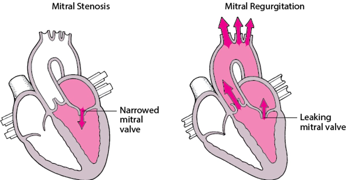
Understanding Stenosis and Regurgitation
The heart valves can malfunction either by leaking (causing regurgitation) or by not opening adequately and thus partially blocking the flow of blood through the valve (causing stenosis). Stenosis and regurgitation can affect any of the heart valves. These two disorders are shown below affecting the mitral valve. | |
Normally, just after the left ventricle finishes contracting and starts to relax and fill with blood again (during diastole), the aortic valve closes, the mitral valve opens, and some blood flows from the left atrium into the left ventricle. Then the left atrium contracts, ejecting more blood into the left ventricle. | As the left ventricle begins to contract (during systole), the mitral valve closes, the aortic valve opens, and blood is ejected into the aorta. |
In mitral stenosis, the mitral valve opening is narrowed, and blood flow from the left atrium into the left ventricle during diastole is reduced. | In mitral regurgitation, the mitral valve leaks when the left ventricle contracts (during systole), and some blood flows backward into the left atrium. |


Repairing or replacing a heart valve
A faulty valve may be repaired or replaced. Repair may require surgery but may sometimes be accomplished during heart catheterization, particularly when the problem is a valve with stenosis. A stenotic valve can sometimes be stretched open using a procedure called balloon valvuloplasty. In this procedure, a catheter with a balloon at the tip is threaded through a vein or artery into the heart. Once across the faulty valve, the balloon is inflated, separating the valve cusps. This procedure does not require a general anesthetic and allows a quick recovery.
Two types of valves are available for replacement
Mechanical type
Bioprosthetic type (made from the heart valve of a pig or cow)
Overview of Hemorrhagic Stroke). Bioprosthetic valves generally deteriorate and require replacement after 10 to 12 years but do not require use of anticoagulants for more than 3 to 6 months after surgery, but people with bioprosthetic valves will need to take aspirin also to help prevent blood clots. Some bioprosthetic valves are more durable.
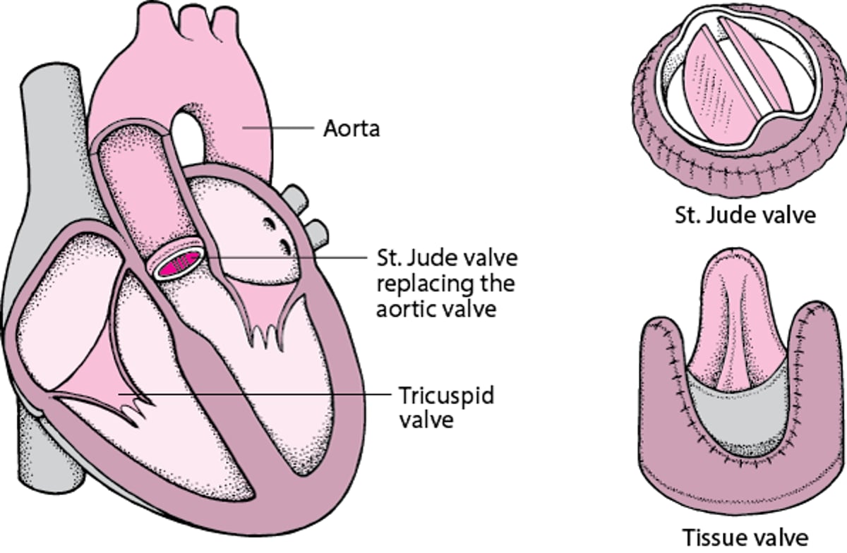
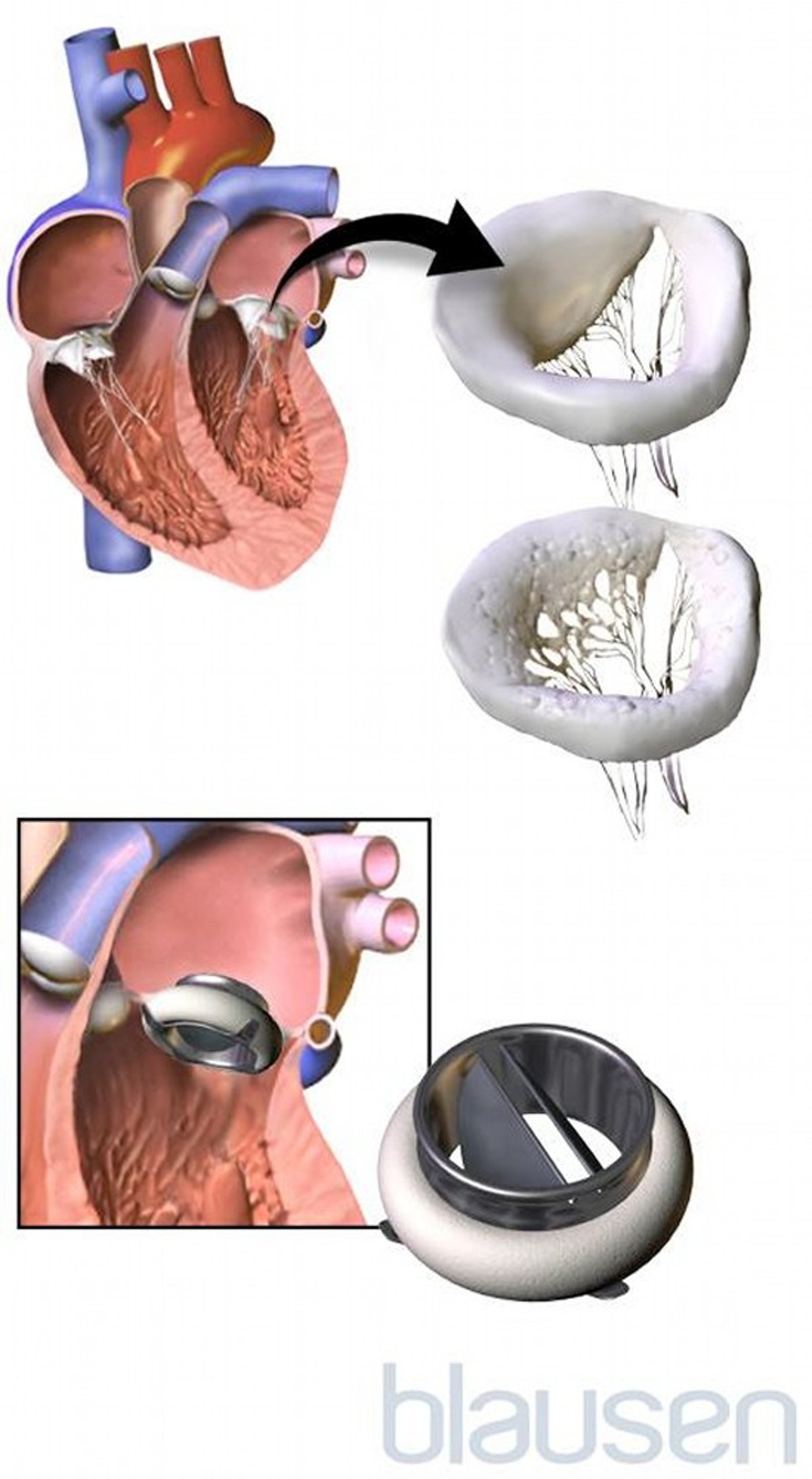
Replacing a Heart Valve
A damaged heart valve may be replaced with a mechanical valve made of plastic and metal or with a bioprosthetic valve made of heart valve tissue, usually from pigs, placed in a synthetic ring. There are many types of mechanical valves. A St. Jude valve is commonly used. Choice of a valve depends on many factors, including characteristics of the valve. A mechanical valve lasts longer than a bioprosthetic valve but requires that anticoagulants be taken indefinitely to prevent the formation of blood clots on the valve. A bioprosthetic valve requires only short-term use of anticoagulants. So whether a person can take anticoagulants is an important factor. For example, anticoagulants may not be appropriate for women of childbearing age because anticoagulants cross the placenta and may affect the fetus. Also considered are
When an aortic valve is being replaced, a mechanical valve is usually chosen for people who are under age 50 and a bioprosthetic valve is chosen for those age 50 or older. When a mitral valve is being replaced, a mechanical valve is usually chosen for people who are under age 65 and a bioprosthetic valve is chosen for those age 65 or older. The tricuspid and pulmonic valves do not need to be replaced or repaired as often as the aortic or mitral valves. For heart valve replacement, a general anesthetic is given. The heart must be still to be operated on, so a heart-lung machine is used to pump blood through the bloodstream. The damaged valve is removed, and the replacement valve is sewn in place. The incisions are closed, the heart-lung machine is disconnected, and the heart is restarted. The operation takes from 2 to 5 hours. For some people, a heart valve can be replaced using a less invasive procedure (without cutting through the sternum), available at some medical centers. The length of the hospital stay varies from person to person. Full recovery may take 6 to 8 weeks. |


Abnormal valves and all replacement valves can become infected. People with replacement valves need to take prophylactic antibiotics, which are antibiotics taken at certain times (for example, before some dental or medical procedures)r to prevent bacterial infection of the valves (infective endocarditis).
Also, blood clots may form on replacement valves. The blood clots may partially block the valve or break loose and travel through the bloodstream to block arteries elsewhere in the body (for example, causing a stroke).
Doctors use echocardiography to monitor replaced valves by looking at the blood flow through the valves and the rest of the heart.
More Information
The following English-language resource may be useful. Please note that THE MANUAL is not responsible for the content of this resource.
American Heart Association: Heart Valve Disease: Provides comprehensive information on diagnosis and treatment of diseases of the heart valves