

One or more bones of the pelvis may be broken. These fractures range from a small chip of bone being broken off, to fractures due to slight force (as can occur in older adults with osteoporosis), to fractures due to great force (as occurs in car crashes).
Most pelvic fractures cause considerable pain, even when people are sitting or lying down.
Severe pelvic fractures can result in life-threatening bleeding and may be accompanied by serious injuries to other organs.
X-rays can show most pelvic fractures, but computed tomography is usually also done.
Minor fractures require only pain relievers, but more severe fractures must be stabilized with an external device or with plates and screws inserted surgically.
(See also Overview of Fractures.)
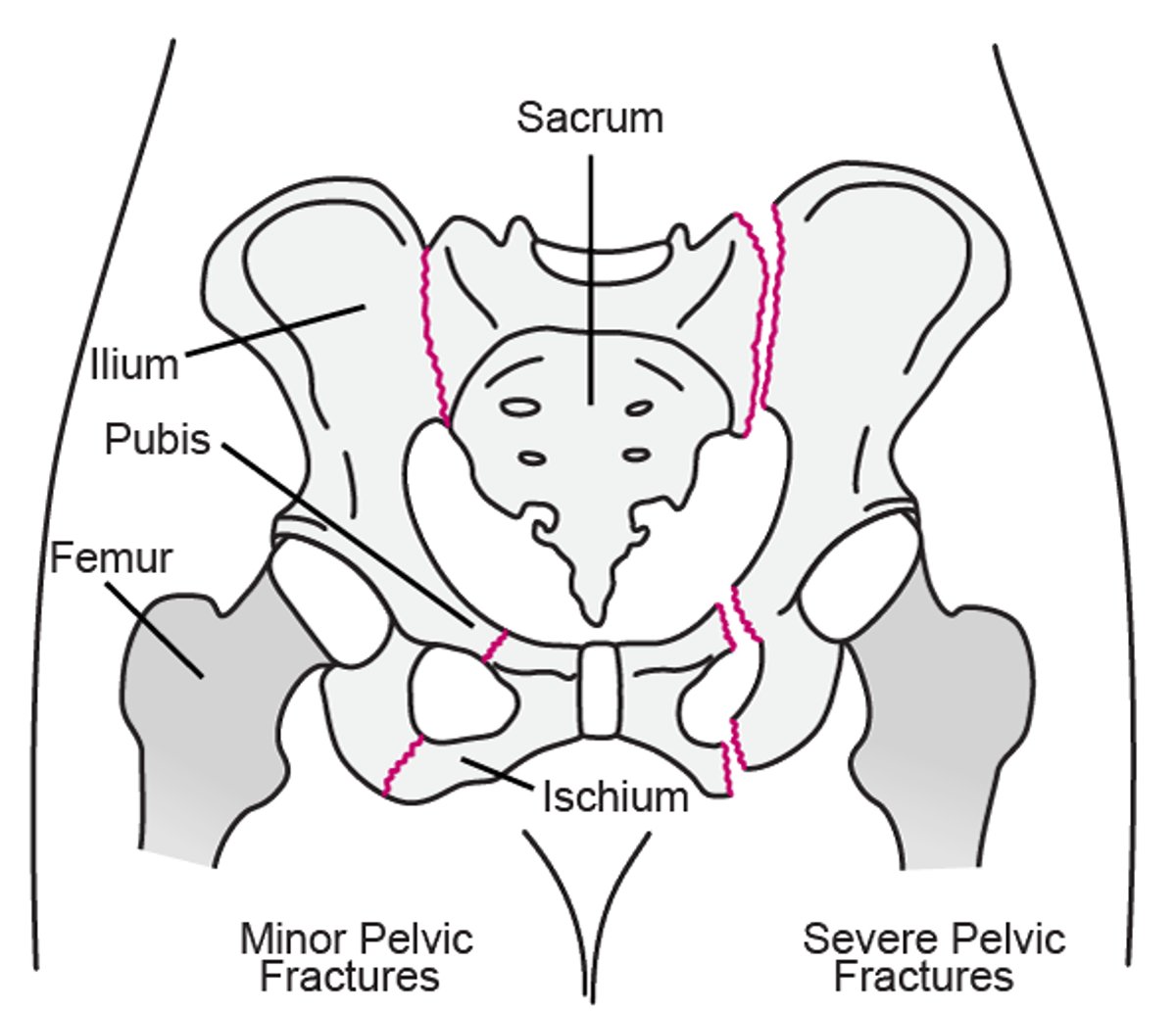
The pelvis, located at the bottom of the torso, is made up of three bones:
Ilium, the largest and uppermost bone of the pelvis, located in the back
Pubis, the middle bone of the pelvis, located in the front
Ischium, the bottom bone of the pelvis, located in the back
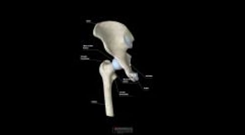
The pelvic bones form the socket for the top of the thighbone (femur) and, with the thighbone, form the hip joint. The pelvis is attached to the tailbone (sacrum) by ligaments at the base of the spine. Many ligaments hold these bones in place. Muscles of the thigh (hamstring and gluteal muscles) are attached to the pelvis by tendons.
Pelvic Fractures
Fractures (shown in red below) may occur in the ilium, pubis, or ischium bones. |

Causes of Pelvic Fractures
In young adults, severe fractures of the entire pelvis can result from high-speed car or motorcycle crashes, collision of a car with a pedestrian, or falls from a height. These fractures can cause life-threatening bleeding, whether the skin is broken or not. Dangerously low blood pressure (shock) can result. Also nearby nerves and organs, such as the bladder, reproductive organs, and intestine, may be damaged. Severe fractures are usually unstable.
In some older adults, osteoporosis weakens the bones. Thus, they may fracture part of the pelvis if they fall—for example, when getting out of the bathtub, when going down steps, or even while standing on level ground.
One type of pelvic fracture tends to occur mainly in adolescents, especially those involved in sports. These fractures (called avulsion fractures) usually occur when a muscle contracts suddenly and pulls a small piece of bone from the ischium where the hamstring muscles are attached (near the base of the buttocks).
Symptoms of Pelvic Fractures
Most fractures of the pelvis cause considerable pain in the groin, even when people are lying down or sitting. The pain becomes much worse when people try to walk, although some can walk. The area is often swollen and bruised. People may try to keep the hip or knee bent in a specific position to avoid making the pain worse.
If pelvic fractures are severe and unstable, people have extreme pain and cannot walk. However, some minor fractures are much less painful.
If other structures are injured, people may have other symptoms. They may have blood in their urine, difficulty urinating, uncontrollable loss of urine (incontinence), or bleeding from the rectum or vagina.
If the socket of the hip joint has been damaged, people may be permanently disabled.
When bleeding is severe enough to cause shock or when other organs are damaged, people may die.
Diagnosis of Pelvic Fractures
X-rays
Usually computed tomography
Physical examination to check for other injuries
(See also Diagnosis of Fractures.)
If people think they have injured their pelvis, they should go to an emergency department. If people are in a major accident, they are usually taken to an emergency department by ambulance. There, injuries are identified and treatment is begun.
Doctors suspect a pelvic fracture when people have pain in the groin or have had a major injury. X-rays are then taken. X-rays can show most pelvic fractures. Computed tomography (CT) is usually done to identify all the broken bone fragments and to check for other injuries.
To check for other injuries (particularly urinary tract injuries), doctors do a physical examination. It includes a neurologic examination, a digital rectal examination, and, in women, a pelvic examination. Urine tests are also done. If doctors suspect injury to the urinary tract, CT or another imaging test of the urinary tract is done.
Treatment of Pelvic Fractures
For minor, stable fractures, usually only pain relievers and walking
For severe fractures, an external device or surgery to immobilize the pelvis
If needed, procedures to stop bleeding
When people who have been in a major accident are taken to an emergency department, serious injuries must be treated as soon as possible. If bleeding is severe, steps must be taken immediately to stop the bleeding. People usually need to be admitted to a hospital.
Minor, stable pelvic fractures typically heal without causing permanent disabilities. Surgery is rarely needed, but bed rest may be needed. However, bed rest should be for as short a time as possible. Pain relievers (analgesics) can help relieve pain enough so that people can walk. To avoid the weakness, stiffness, and other complications that occur with bed rest, people should walk, stand, and put their full weight on the joint as soon as possible, even if they can do so only for a short time. Trying to walk does not injure the area further. Most people can walk short distances without a walker by 1 week and can walk without aid and with only mild discomfort in 1 to 2 months.
Severe pelvic fractures, which are often unstable, must be immobilized. Emergency personnel usually stabilize the joint by wrapping it with strips of cloth or with a binder designed for this purpose until the injury can be more permanently stabilized. To more securely stabilize the injury, especially if there are other serious injuries, doctors may attach a rigid metal frame to the pelvis, outside the body, using long screws inserted through the skin into the bones. This device is called an external fixator. Once the injury is more stable, surgery is usually done to align the broken pieces of bone and to insert plates and screws to hold them in place (called open reduction with internal fixation, or ORIF). After the pelvis is stabilized, people are encouraged to walk as soon as possible. Sometimes the fracture is surgically repaired immediately, without use of an external fixator.
If bleeding continues, embolization or pelvic packing may be done:
Embolization involves inserting a thin, flexible tube (catheter) into a blood vessel and threading it to the injured blood vessel. Tiny coils or a gel-like substance is inserted through the catheter into the bleeding blood vessels to block them and thus stop the bleeding.
Pelvic packing involves surgery to insert material around the damaged organs in the pelvis. This material absorbs the blood and puts pressure on blood vessels, which helps slow or stop the bleeding. Once the bleeding has stopped, usually days later, surgery is done to remove the packing and the pelvis is repaired surgically (ORIF).
Other injuries are treated as needed.


