

The retina is the light-sensitive structure at the back of the eye. Blunt injury may cause part of the retina or the entire retina to tear or to separate (detach) from its underlying surface at the back of the eyeball.
(See also Overview of Eye Injuries.)
Usually, only part of the retina detaches (often the outside edge, or peripheral part, of the retina), but if treatment does not occur shortly after the injury, more of the retina can detach. When the retina detaches, it separates from part of its blood supply. Unless the retina is reattached soon, it may be permanently damaged by lack of blood.
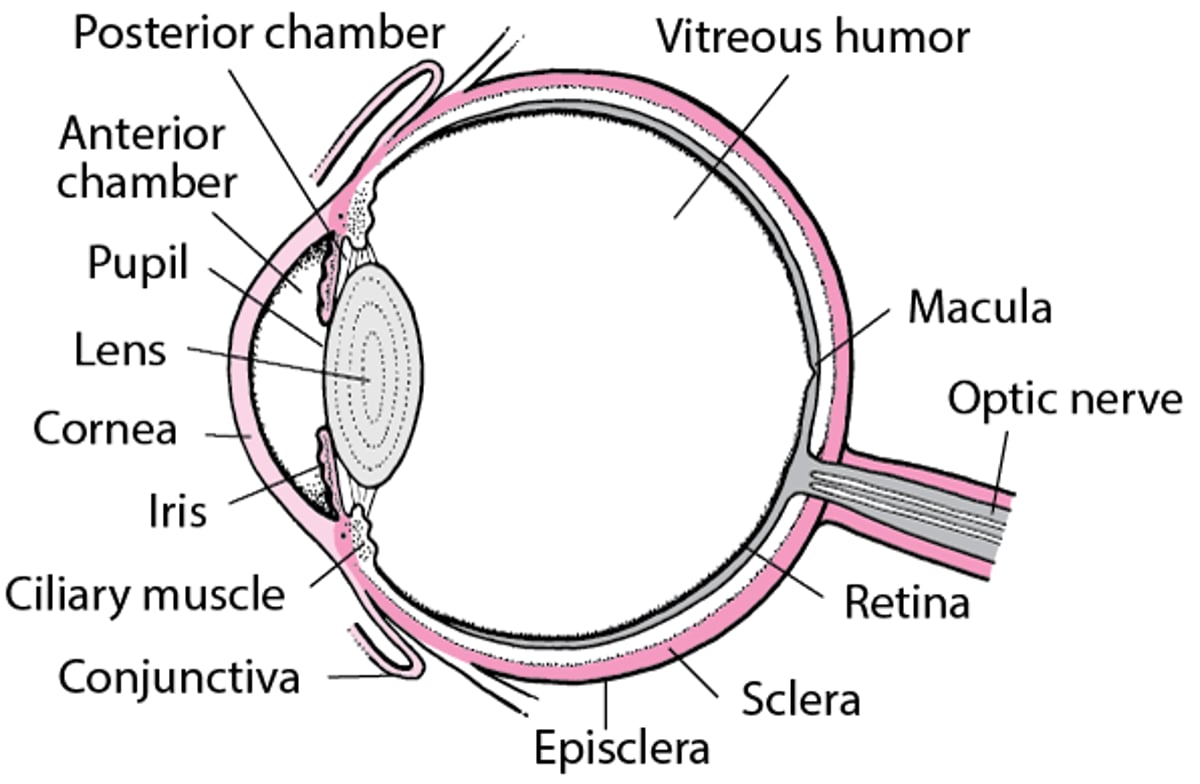
An Inside Look at the Eye

Symptoms of Traumatic Retinal Detachment
A retinal detachment is painless and may be asymptomatic initially or if there is a small detachment. People usually have the following symptoms, which worsen as more of the retina detaches:
Seeing an increase in floating shapes (floaters—spots/lines that appear to move through a person's field of vision)
Seeing many flashes of bright light that last less than a second (photopsias)
Having blurred vision
Having an area of their vision where they cannot see (a visual field cut)
Peripheral vision is typically lost first, and vision loss spreads as the detachment progresses. The loss of vision causes grayness in the field of vision or resembles a curtain or veil falling across the line of sight. People may have blood in the jellylike vitreous humor near the back of the eye (vitreous hemorrhage). If the macula becomes detached, vision rapidly deteriorates, and everything becomes blurred. A person with any of these symptoms needs to see a doctor as soon as possible.
Diagnosis of Traumatic Retinal Detachment
A doctor's examination of the eye
Sometimes ultrasonography
An ophthalmologist (a medical doctor who specializes in the evaluation and treatment—surgical and nonsurgical—of eye disorders) examines the back of the eye with a bright light (ophthalmoscopy) after applying eye drops to dilate the pupil. Sometimes an ultrasound examination is done.
Treatment of Traumatic Retinal Detachment
Surgery
Most retinal detachments can be surgically repaired. The surgeon seals retinal tears with laser surgery or freezing therapy (cryotherapy). For large retinal detachments, the surgeon may bring the retina and the wall of the eye together, either by placing a silicone band around the eye (called a scleral buckle) or by removing the vitreous jelly behind the lens and in front of the retina with surgery called a vitrectomy. A gas bubble is often used to hold and flatten the retina in place. For small detachments, the retina can be reattached using laser surgery or cryotherapy plus a gas bubble (called pneumatic retinopexy). Treatment should be completed soon after the detachment to give the best possible vision.


