

A submandibular space infection is a bacterial infection of the tissues below the mouth.
Submandibular space infection can cause pain and tenderness under the tongue and/or under the jaw, and swelling that may block the airway
Doctors can usually diagnose submandibular space infection by examining the mouth, but computed tomography is sometimes needed.
Doctors insert a breathing tube to keep the airway open and give antibiotics.
Bacteria can spread from an infected lower tooth to the tissue under and around the tongue. People with poor dental hygiene and people who have had a tooth pulled or a jaw fracture are at higher risk. The infection causes swelling that can block the airway causing difficulty breathing and sometimes death.

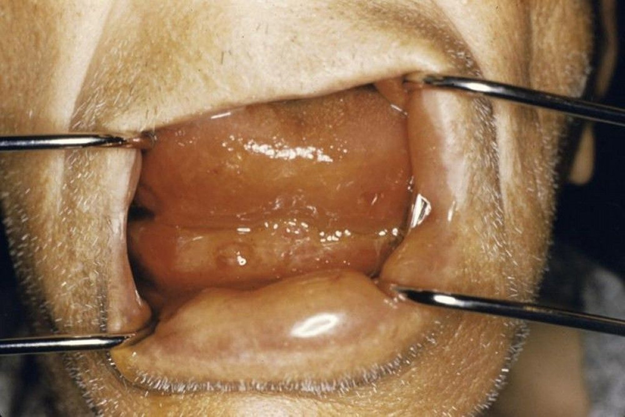
Ludwig angina can cause swelling below the tongue that can block the airway.
Image provided by Clarence T. Sasaki, MD.
Symptoms of Submandibular Space Infection
People with a submandibular space infection have pain and tenderness under the tongue and/or under the jaw. The pain is worse with opening the mouth or swallowing.
Fever and chills are common.
Later, swelling worsens, which may cause drooling, and people may make a sound (for example, squeaking or gasping) when they breathe in (called stridor). Once swelling occurs, blockage of the airway and death may occur within hours.
Diagnosis of Submandibular Space Infection
A doctor's evaluation
Sometimes computed tomography
Doctors usually can diagnose submandibular space infection by examining the mouth.
If the examination is unclear, computed tomography (CT) is done. However, if an airway blockage appears to be developing or may occur soon, treatment is started quickly, and CT is postponed.
Treatment of Submandibular Space Infection
A breathing tube followed by surgery to drain the infected area
Antibiotics
Treatment for submandibular space infection must be done quickly to prevent blockage of the airway.
Doctors take the person to the operating room and use a thin, flexible viewing (fiberoptic) tube to help guide a plastic breathing tube through the nose into the windpipe (trachea) to keep the airway open. Then doctors surgically open the infected area to allow the infection to drain.
If a breathing tube cannot be placed, a tracheotomy is done.
Antibiotics such as ceftriaxone and clindamycin are given by vein.Antibiotics such as ceftriaxone and clindamycin are given by vein.
Drug Information for the Topic


