Chronic otitis media is defined variously as persistent middle ear effusion for more than three months or as recurrent ear drainage in the setting of a perforated tympanic membrane, also known as chronic suppurative otitis media. Generally accepted criteria for chronic otitis media are three infections in six months or four in twelve months. Globally, it is estimated that chronic otitis media affects 31 million people per year, about one fifth of whom are children younger than five years of age (1).
Chronic otitis media can cause hearing loss and ear drainage. Complications include progressive damage to the middle ear structures as a result of aural polyps, cholesteatoma, and other infections.
Chronic otitis media in children is concerning because of the well-known, long-term, detrimental effects of hearing loss on linguistic, cognitive and psychosocial development (2). Guidelines by the American Academy of Pediatrics state that “otitis media may be related to difficulties in speech and reading, delayed response to auditory input, limited vocabulary, and disturbances in attention.”
Diagnosing and treating cholesteatomas
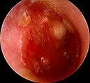
A cholesteatoma is a cyst that forms in the middle ear, mastoid, or epitympanum after chronic otitis media. Although technically benign, it is locally invasive and can damage middle ear structures. Cholesteatomas are sometimes seen during otoscopy as a white mass behind the ear drum.
Because cholesteatoma can result in permanent hearing loss and other serious sequelae such as intracranial abscess, physicians should be on the lookout for this complication, particularly when otorrhea does not stop despite culture-directed antimicrobial treatment. In these cases, imaging or examination under anesthesia is warranted if routine otoscopy is not suspicious.
A CT scan is usually needed to determine the full extent of the cholesteatoma, and an audiogram is typically required to determine the cyst’s impact on hearing.
It’s important to note that cholesteatomas will continue to grow at a slow rate if not removed. In a space as small as the middle ear, even this slow growth can have devastating consequences if not managed appropriately. It’s best to limit the damage by removing a cholesteatoma and reconstructing the middle ear for hearing preservation, if possible.
Hearing reconstruction may be needed if the surgical removal of the cholesteatoma requires sacrifice of the ossicles (bones of hearing) due to the lesion involving these structures.
Talking to patients about chronic otitis media
When talking to patients about chronic otitis media and its complications, it is critical to set realistic expectations regarding the extent of disease, and the likely response to treatment. When discussing cholesteatomas, clinicians should emphasize that although the growth is not malignant, it still can cause serious damage to the ear, or beyond. Clinicians can also refer patients to the Manuals consumer pages on chronic otitis media.
References
- Monasta L, Ronfani L, Marchetti F, et al: Burden of disease caused by otitis media: systematic review and global estimates. PLoS ONE 7: e36226, 2012. doi.org/10.1371/journal.pone.0036226
- Bennett KE, Haggard MP, Silva PA, et al: Behavior and developmental effects of chronic otitis media with effusion into the teens. Archives of Disease in Childhood 85:91–95, 2001. doi: 10.1136/adc.85.2.91