Similar to the motor examination, the sensory examination is designed to localize dysfunction and help determine whether the problem is in the cerebral cortex, thalamus, sensory pathways in the brain or spinal cord, or peripheral nerves.
For localization, the most useful elements of the examination include tests for pain and temperature sensations, which are transmitted through the spinothalamic tract, and position and vibration sensations, which are transmitted through the dorsal column via the medial lemniscus pathway.
Touch can be tested to screen for abnormalities but is not useful for localization, which requires more detailed testing of other types of sensation in different areas of the body.
For the ability to sense a sharp object, the best screening test uses a safety pin or other sharp object to lightly prick the face, torso, and 4 limbs; the patient is asked whether the pinprick feels the same on both sides and whether the sensation is dull or sharp. The sharp object is discarded after use to avoid potential transmission of bloodborne disorders (eg, HIV infection, hepatitis).
Cortical sensory function is evaluated by asking the patient to identify a familiar object (eg, coin, key) placed in the palm of the hand (stereognosis) and numbers written on the palm (graphesthesia) and to distinguish between 1 and 2 simultaneous, closely placed pinpricks on the fingertips (2-point discrimination).
Another indicator of impaired cortical sensory function is extinction, which is inability to identify a stimulus on one side when both sides of the body are tested simultaneously (double simultaneous stimulation) in a patient who can identify the stimulus when one side of the body is tested at a time. For example, when extinction is present, patients report feeling sensation on only one side when simultaneously touched on both sides even though they can feel sensation on both sides when one side is tested at a time.
Temperature sense is usually tested with a cold tuning fork.
Joint position sense is tested by moving the terminal phalanges of the patient’s fingers, then the toes, up or down a few degrees. If the patient cannot identify these tiny movements with eyes closed, larger up-and-down movements are tried before testing the next most proximal joints (eg, testing the ankles if toe movement is not perceived).
Pseudoathetosis refers to involuntary writhing, snakelike movements of a limb that result from severe loss of position sense; motor pathways, including those of the basal ganglia, are preserved. The brain cannot sense where the limb is in space so the limb moves on its own, and the patient must use vision to control the limb’s movements. Typically, when the eyes are closed, the patient cannot locate the limb in space.
Inability to stand with feet together and eyes closed (Romberg test) indicates impaired position sense in the lower extremities. When cerebellar disease is present, the patient tries to stand with the feet apart but as close together as possible without falling and only then closes the eyes. Rarely, a positive result is due to severe bilateral loss of vestibular function (eg, aminoglycoside toxicity).
To test vibration sense, the examiner places a finger under the patient’s distal interphalangeal joint and presses a lightly tapped 128-cycle tuning fork on top of the joint. The patient should note the end of vibration about the same time as the examiner, who feels it through the patient’s joint.
Light touch is tested with a cotton wisp.
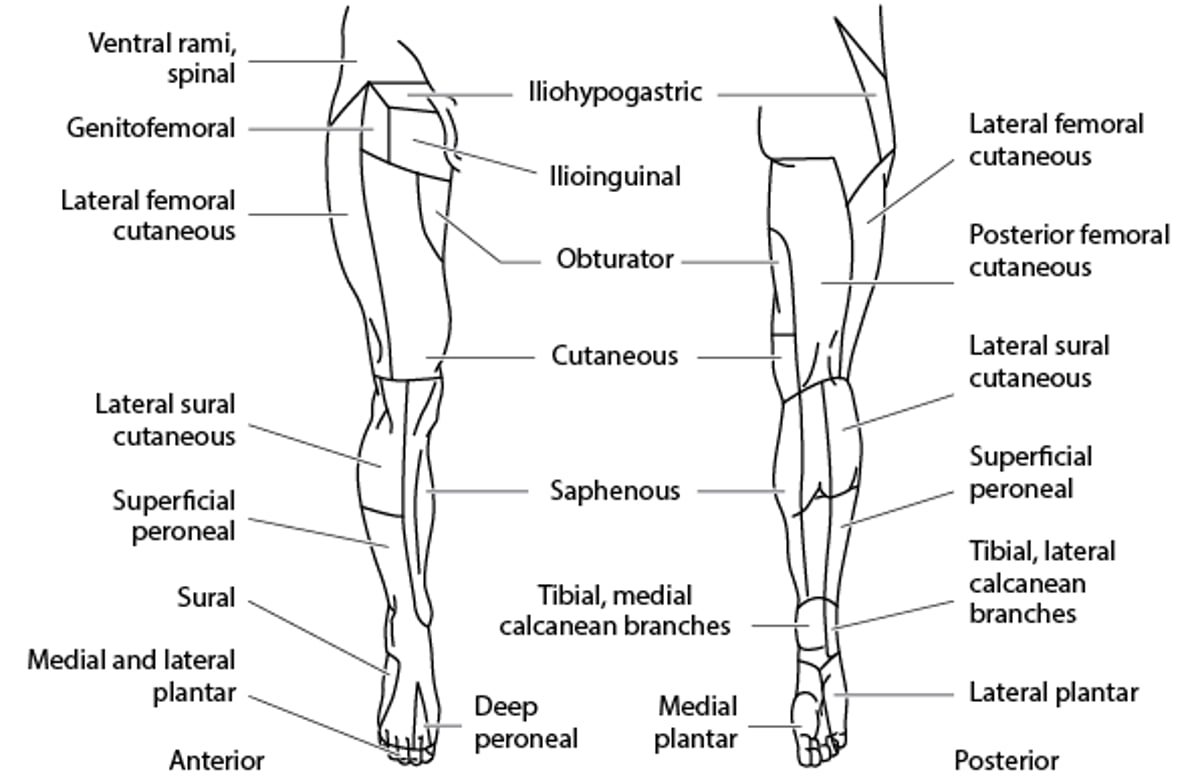
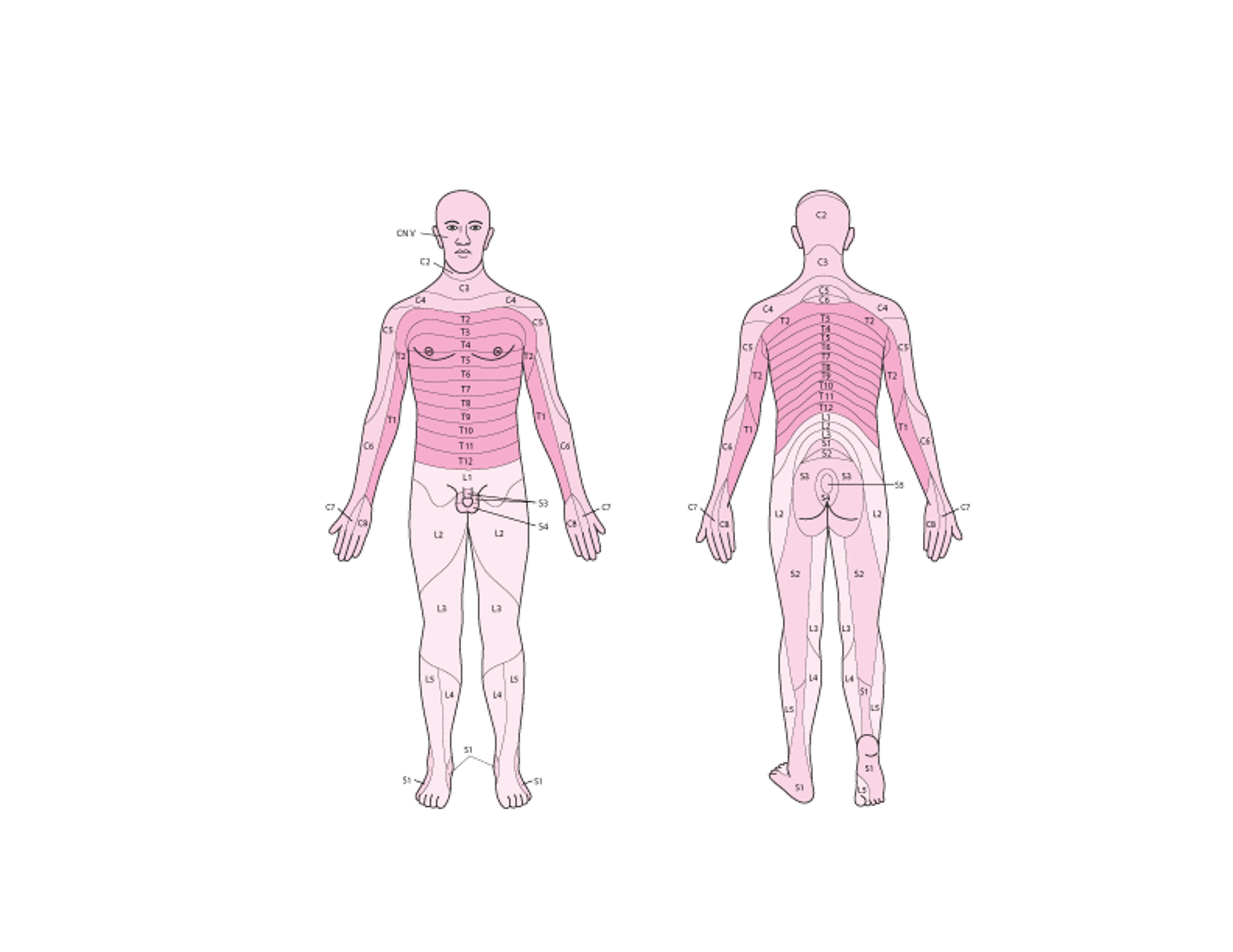
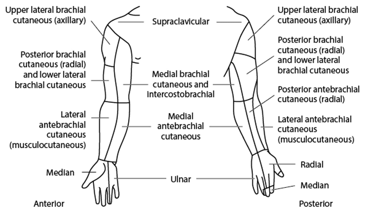
If sensation is impaired, the anatomic pattern suggests location of the lesion (see also figures Sensory Dermatomes, Cutaneous Nerve Distribution: Upper Limb, and Cutaneous Nerve Distribution: Lower Limb):
Stocking-glove distribution: Distal peripheral nerves
Single dermatomal or nerve branch distribution: Isolated nerves (mononeuritis multiplex) or nerve roots (radiculopathy)
Patchy sensory, motor, and reflex deficits in a limb: Brachial or pelvic plexus
Sensation reduced below a certain dermatomal level: Spinal cord
Saddle area sensory loss: Cauda equina
Crossed face-body pattern: Brain stem
Hemisensory loss: Brain
Midline hemisensory loss: Thalamus or functional (psychiatric)
Location of the lesion is confirmed by determining whether motor weakness and reflex changes follow a similar pattern.
(See also Introduction to the Neurologic Examination.)
Sensory Dermatomes

Cutaneous Nerve Distribution: Upper Limb
(Redrawn from Anatomy,ed. 5, edited by R O’Rahilly. Philadelphia, WB Saunders Company, 1986; used with permission.) |

Cutaneous Nerve Distribution: Lower Limb
(Redrawn from Anatomy,ed. 5, edited by R O’Rahilly. Philadelphia, WB Saunders Company, 1986; used with permission.) |