A corneal ulcer is a corneal epithelial defect with underlying inflammation usually due to invasion by bacteria, fungi, viruses, or Acanthamoeba
(See also Introduction to Corneal Disorders.)
Etiology of Corneal Ulcer
Corneal ulcers have many causes (see table Causes of Corneal Ulcers). Herpes simplex keratitis is discussed separately.
Bacterial ulcers are most commonly due to contact lens wear and are rarely due to secondary infection from traumatic abrasion or herpes simplex keratitis. The response to the treatment depends mostly on the bacterial species, and the ulcer may be particularly refractory to treatment.
The time course for ulcers varies. Ulcers caused by Acanthamoeba (also most commonly due to exposure to contaminated water while wearing contact lenses) and fungi (most commonly due to trauma with vegetable material) are indolent but progressive, whereas those caused by Pseudomonas aeruginosa (seen most frequently in contact lens wearers) develop rapidly, causing deep and extensive corneal necrosis. Wearing contact lenses while sleeping or wearing inadequately disinfected contact lenses can cause corneal ulcers (see Contact Lenses: Care and Complications).
Pathophysiology of Corneal Ulcer
Ulcers are characterized by corneal epithelial defects with underlying inflammation and necrosis of the corneal stroma. Corneal ulcers tend to heal with scar tissue, resulting in opacification of the cornea and decreased visual acuity. Uveitis, corneal perforation with iris prolapse, pus in the anterior chamber (hypopyon), panophthalmitis, and destruction of the eye may occur without treatment and, on occasion, even with the best available treatment, particularly if treatment is delayed. More severe symptoms and complications tend to occur with deeper ulcers.
Symptoms and Signs of Corneal Ulcer
Conjunctival redness, eye ache, foreign body sensation, photophobia, and lacrimation may be minimal initially.

Corneal ulcers due to Acanthamoeba are often intensely painful and may show transient corneal epithelial defects, multiple corneal stromal infiltrates, and, later, a large ring-shaped infiltrate. Fungal ulcers, which are more chronic than bacterial ulcers, are densely infiltrated and show occasional multiple discrete islands of infiltrate (satellite lesions) at the periphery. Dendritic ulcers are characteristic of herpes simplex keratitis.
Diagnosis of Corneal Ulcer
Slit-lamp examination

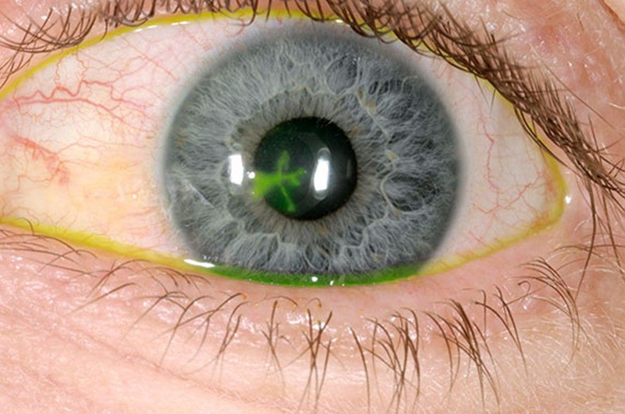
DR P. MARAZZI/SCIENCE PHOTO LIBRARY
Diagnosis is made by slit-lamp examinationAcanthamoeba.
Treatment of Corneal Ulcer
Initially empiric topical broad-spectrum antibiotic therapy
More specific antimicrobial therapy directed at the cause
FusariumCandida
If Acanthamoeba
Key Points
Causes of corneal ulcers include infection of the cornea (including overwearing of contact lenses), eye trauma, abnormalities of the eyelid, and nutritional deficiencies.
Ulcers may be accompanied by circumcorneal hyperemia and WBC layering in the anterior chamber (hypopyon).
All but the smallest ulcers are cultured, usually by an ophthalmologist.
Treatment usually involves frequent (eg, every 1 to 2 hours around the clock) application of topical antimicrobials.