A pheochromocytoma is a tumor that usually originates from the chromaffin cells of the adrenal glands, causing overproduction of catecholamines, powerful hormones that induce high blood pressure and other symptoms.
High blood pressure is the most important symptom, but a fast and pounding pulse, excessive sweating, light-headedness when standing, rapid breathing, severe headaches, and many other symptoms may also occur.
Doctors measure the blood levels of catecholamines or products created when catecholamines are broken down by the body and use imaging tests to try to find the tumor.
Usually, the best treatment is to remove the pheochromocytoma.
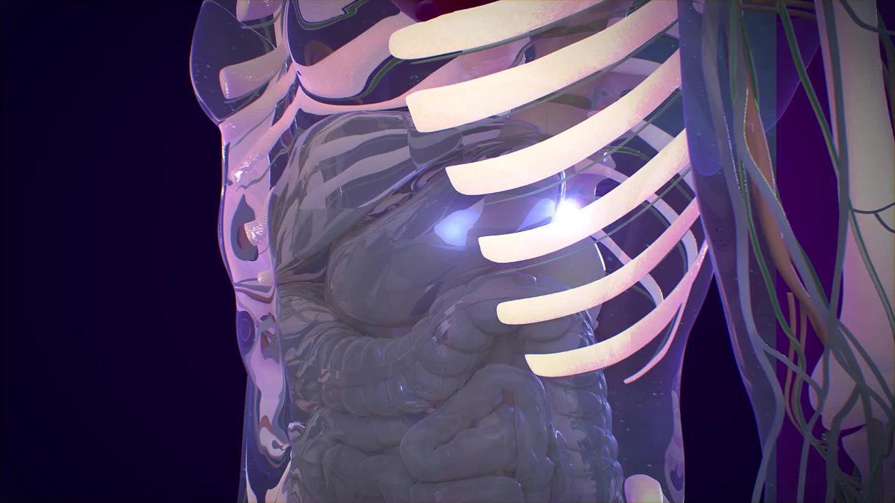
(See also Overview of the Adrenal Glands and the figure A Close Look at the Adrenal Glands.)
Most pheochromocytomas grow within the inner part of the adrenal glands (the medulla). About 10% grow in chromaffin cells outside the adrenal glands. Less than 10% of pheochromocytomas that grow within the adrenal glands are cancerous, but this percentage is higher for those outside the adrenal glands. Pheochromocytomas are most common in people between the ages of 20 and 40 years.
Some people who develop pheochromocytomas have a rare inherited condition called multiple endocrine neoplasia that makes them prone to tumors in the thyroid, parathyroid, and adrenal glands. Pheochromocytomas may also develop in people who have von Hippel–Lindau disease and in those who have neurofibromatosis (von Recklinghausen disease) or a number of other genetic diseases. It is likely that nearly 50% of people who have pheochromocytomas have a genetic or familial disease such as these.
Symptoms of Pheochromocytoma
Pheochromocytomas may be quite small. However, even a small pheochromocytoma can produce large amounts of potent catecholamines. Catecholamines are hormones such as adrenaline (epinephrine), norepinephrine, and dopamine, which tend to greatly increase blood pressure and heart rate and cause other symptoms usually associated with life-threatening situations.
The most prominent sign of a pheochromocytoma is high blood pressure, which may be very severe. However, only about 1 in 1,000 people with high blood pressure has a pheochromocytoma. Symptoms include
A fast and pounding heart rate
Excessive sweating
Light-headedness when standing
Rapid breathing
Cold and clammy skin
Severe headaches
Chest and stomach pain
Nausea and vomiting
Vision disturbances
Tingling fingers
Constipation
An odd sense of impending doom
When these symptoms appear suddenly and forcefully, they can feel like a panic attack.
In half of affected people, symptoms come and go, sometimes triggered by pressure on the tumor, massage, medications (especially anesthetics and beta-blocking medications), emotional trauma, and, on rare occasions, the simple act of urination. However, many people may have these symptoms as manifestations of an anxiety state, not a glandular disorder.
Diagnosis of Pheochromocytoma
Blood and urine tests
Computed tomography or magnetic resonance imaging
Doctors may not suspect a pheochromocytoma, because almost half of the people have no symptoms other than persistent high blood pressure. However, when high blood pressure occurs in a young adult, comes and goes, or accompanies other symptoms of pheochromocytoma, doctors may request certain laboratory tests. For example, the level of certain catecholamines or products created when these catecholamines are broken down may be measured in blood or urine samples.
Because of high blood pressure and other symptoms, doctors may prescribe a beta-blocker before knowing that the cause is a pheochromocytoma. Beta-blockers can make high blood pressure worse in people with pheochromocytoma. This paradoxical reaction often makes the diagnosis of pheochromocytoma clear.
If the level of catecholamines is high, computed tomography (CT), magnetic resonance imaging (MRI), or another imaging test can help locate the pheochromocytoma. A test using injected radioactive chemicals that tend to accumulate in pheochromocytomas can also be useful. A scan is then done to see where the radioactive chemicals are.
Once pheochromocytoma has been confirmed, doctors typically do genetic tests on the person.
Treatment of Pheochromocytoma
Surgery to remove the tumor
Medications to control blood pressure