

The chest (thoracic) cavity is the area surrounded by the thoracic spine (vertebrae), the ribs, the breastbone (sternum), and the diaphragm. The lungs are housed in the chest cavity, a space that also includes the heart, which is part of the mediastinum. (See also Overview of the Respiratory System.)
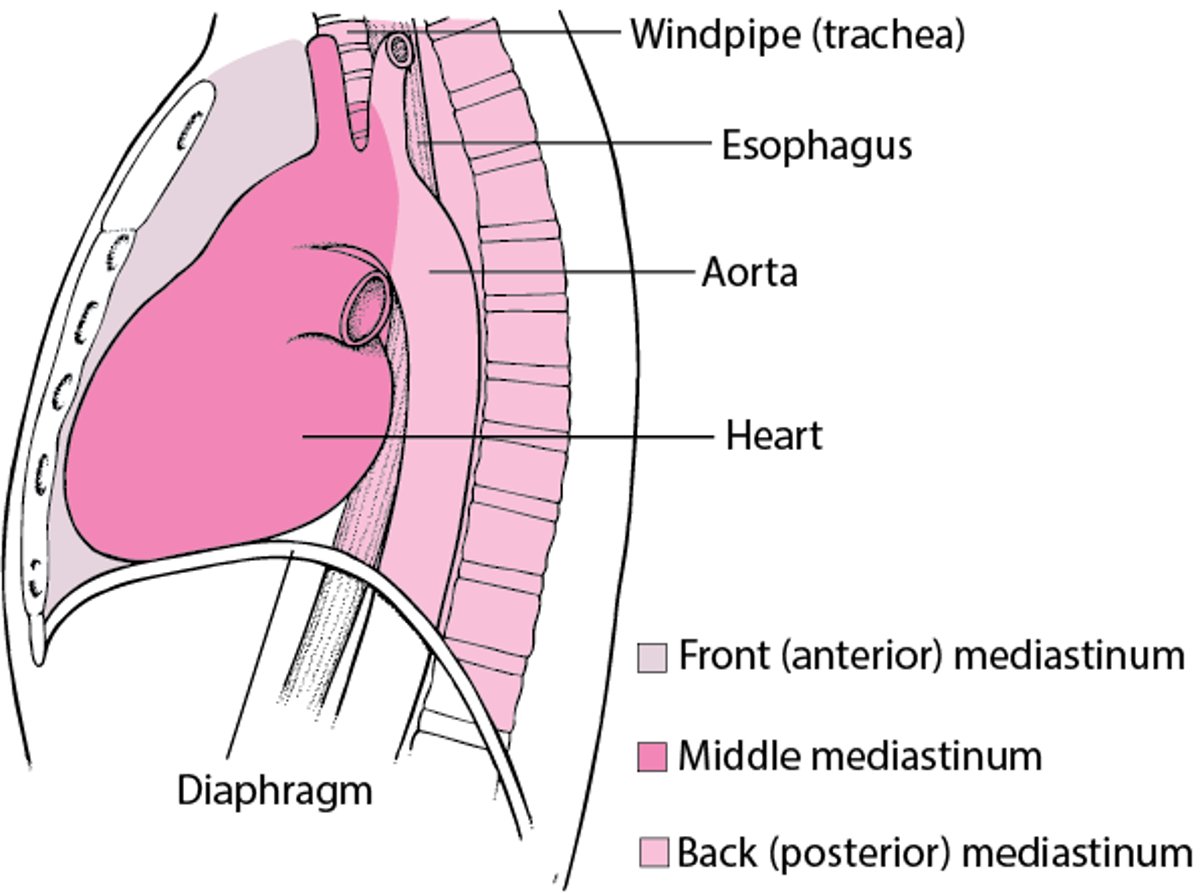
The mediastinum is in the center of the chest and contains the heart, thymus, and lymph nodes, along with portions of the aorta, vena cava, trachea, esophagus, and various nerves. It encompasses the area bordered by the breastbone (sternum) in front, the spinal column in back, the entrance to the chest cavity above, and the diaphragm below. The mediastinum isolates the left and right lung from each other so that they function as 2 separate chest cavities. For example, if the chest wall is punctured on one side, causing the lung on that side to collapse, the other lung remains inflated and functioning, because the 2 lungs are separated by the mediastinum.
Locating the Mediastinum

The rib cage is formed by the sternum, ribs, and spine, protecting the lungs and other organs in the chest. The 12 pairs of ribs curve around the chest from the back. Each pair is joined to the bones (vertebrae) of the spine. In the front of the body, the upper 7 pairs of ribs are attached to the sternum by cartilage. The eighth, ninth, and tenth pairs of ribs join the cartilage of the pair above. The last 2 pairs (floating ribs) are shorter and do not join in the front (see figure Diaphragm's Role in Breathing).


