


The liver produces about 500 to 600 mL of bile each day. Bile is isosmotic with plasma and consists primarily of water and electrolytes but also organic compounds: bile salts, phospholipids (mostly lecithin), cholesterol, bilirubin, and other endogenously produced or ingested compounds, such as proteins that regulate gastrointestinal function and drugs or their metabolites. Bilirubin is a degradation product of heme compounds from worn-out red blood cells (RBCs) and is the pigment that gives bile its yellow-green color.
Bile salts (bile acids) are the major organic component in bile. The liver uses active transport to secrete bile salts into the canaliculus, the cleft between adjacent hepatocytes. Canalicular transport is the rate-limiting step in bile formation. Once secreted, bile salts draw other bile components (particularly sodium and water) into the canaliculus by osmosis. Bile salts are also biologic detergents that enable the body to excrete cholesterol and potentially toxic compounds (eg, bilirubin, drug metabolites). The function of bile salts in the duodenum is to solubilize ingested fat and fat-soluble vitamins, facilitating their digestion and absorption. From the liver, bile flows from the intrahepatic collecting system into the right or left hepatic duct, then into the common hepatic duct.
During fasting, about 75% of the bile secreted passes from the common hepatic duct into the gallbladder via the cystic duct. The rest flows directly into the common bile duct (formed by the junction of the common hepatic and cystic ducts) into the duodenum. During fasting, the gallbladder absorbs up to 90% of bile water, concentrating and storing bile.
View of the Liver and Gallbladder

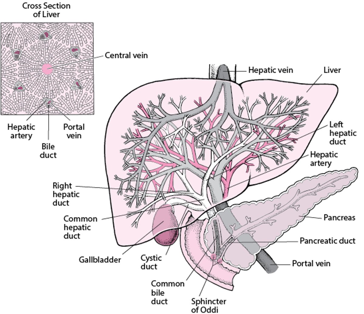
Bile empties from the gallbladder into the common bile duct. The common bile duct joins with the pancreatic duct to form the ampulla of Vater, which empties into the duodenum. Before joining the pancreatic duct, the common bile duct tapers to a diameter of ≤ 0.6 cm.
The sphincter of Oddi, which surrounds both the pancreatic duct and the common bile duct, includes a sphincter for each duct. Bile does not normally flow retrograde into the pancreatic duct. These sphincters are highly sensitive to cholecystokinin and other gut hormones (eg, gastrin-releasing peptide) and to alterations in cholinergic tone (eg, by anticholinergic drugs).
Eating releases gut hormones and stimulates cholinergic nerves, causing the gallbladder to contract and the sphincter of Oddi to relax. As a result, the gallbladder empties 50 to 75% of its contents into the duodenum. Conversely, during fasting, an increase in sphincter tone facilitates gallbladder filling.
Bile salts are poorly absorbed by passive diffusion in the proximal small bowel; most intestinal bile salts reach the terminal ileum, which actively absorbs 90% of bile salts into the portal venous circulation. Returned to the liver, bile salts are efficiently extracted, promptly modified (eg, conjugated if they arrive in the free form), and secreted back into bile. Bile salts circulate through this pathway from liver to gut to liver—the enterohepatic circulation—10 to 12 times/day.
Most disorders of the biliary tract result from gallstones, although acalculous biliary pain occurs in the absence of gallstones and postcholecystectomy syndrome occurs after the gallbladder itself has been removed. Gallstones in the gallbladder (cholelithiasis) are usually asymptomatic. Bile flow may be blocked by gallstones in the bile ducts (choledocholithiasis), triggering biliary colic or causing inflammation of the gallbladder (cholecystitis). Cholecystitis may be acute, developing over hours, or chronic, persisting for a long time.
Blockage of the bile ducts can also cause inflammation, usually with bacterial infection, of the bile ducts (acute cholangitis). Bile flow can be blocked or slowed (called cholestasis) by tumors or, in patients who have AIDS, by strictures caused by opportunistic infections (AIDS cholangiopathy). Cholestasis can also lead to inflammation, fibrosis, and strictures of the bile ducts (called sclerosing cholangitis). Usually, the cause of sclerosing cholangitis is unknown (called primary sclerosing cholangitis [PSC]).


