


Eosinophilic esophagitis is a chronic immune-mediated disease of the esophagus resulting in eosinophil-predominant inflammation of the esophagus; it can cause reflux-like symptoms, dysphagia, and food impaction. Diagnosis is by endoscopy with biopsy. Treatment includes proton pump inhibitors, topical corticosteroids, a biologic, dietary changes, and sometimes esophageal dilation.
(See also Overview of Esophageal and Swallowing Disorders.)
Eosinophilic esophagitis is an increasingly recognized disease that can begin at any time between infancy and young adulthood; it occasionally manifests in older adults. It is more common among males.
The cause of eosinophilic esophagitis is likely an immune response to dietary antigens in patients with genetic susceptibility; environmental allergens may also play a role. Untreated chronic esophageal inflammation ultimately can lead to esophageal narrowing and strictures.
Symptoms of Eosinophilic Esophagitis
Infants and children may present with food refusal, vomiting, weight loss, abdominal pain, and/or chest pain.
In adults, esophageal food impaction is sometimes the first manifestation, and most patients have dysphagia. Symptoms of gastroesophageal reflux disease (GERD), such as heartburn, may occur.
Patients often also have manifestations of other atopic disorders (eg, asthma, eczema, allergic rhinitis).
Diagnosis of Eosinophilic Esophagitis
Endoscopy with biopsy
Sometimes a barium swallow
The typical patient with eosinophilic esophagitis has dysphagia for solids and a history of atopy. The diagnosis of eosinophilic esophagitis is also considered when reflux symptoms fail to respond to acid-suppression therapy. It should also be considered in adults who present with esophageal food impaction or in adults who have noncardiac chest pain.
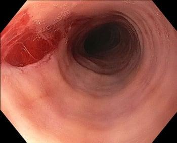
Diagnosis requires endoscopy with biopsy showing eosinophilic infiltration (≥ 15 eosinophils/high-powered field). Although visible abnormalities (eg, linear furrows, strictures, stacked circular rings, loss of vascular markings, white exudates) may be apparent on endoscopy, the appearance can be normal, so biopsies are essential. Because GERD can also cause eosinophilic infiltrates, patients who have mainly reflux symptoms should have biopsies; samples from the proximal and middle esophagus should be processed separately from samples from the distal esophagus.
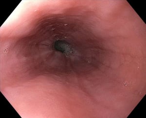
Image provided by Kristle Lynch, MD.
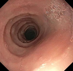
Image provided by Kristle Lynch, MD.

Image provided by Kristle Lynch, MD.

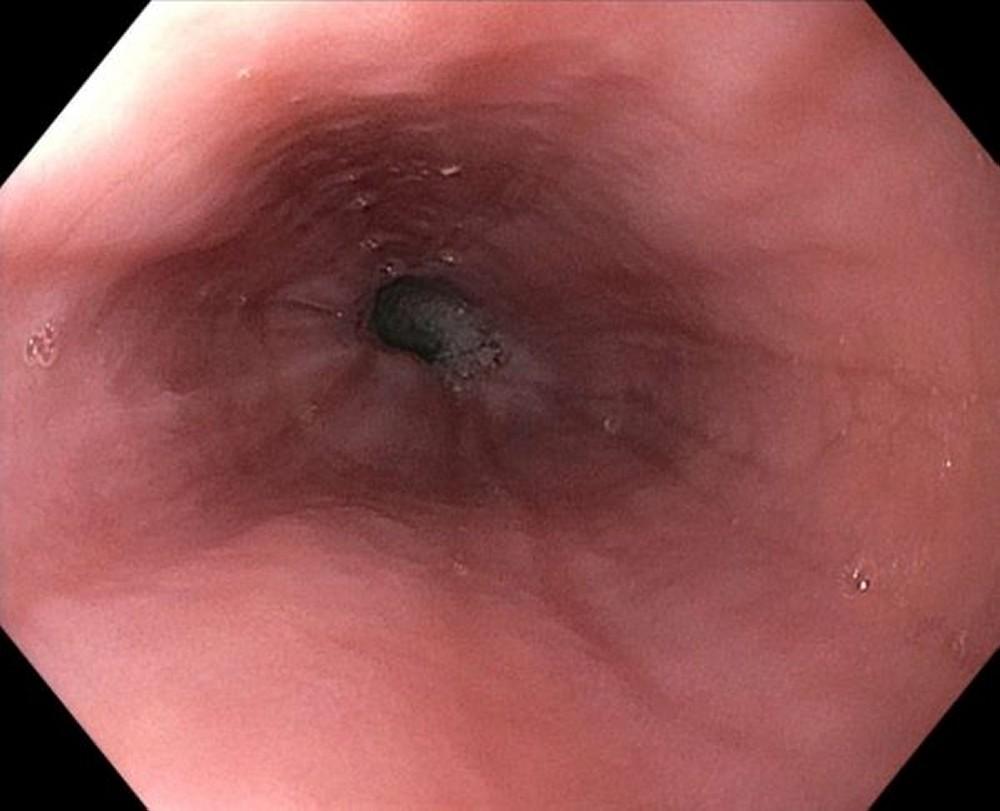
Image provided by Kristle Lynch, MD.

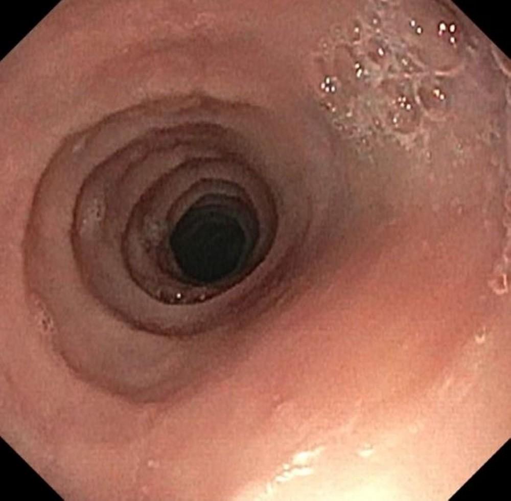
Image provided by Kristle Lynch, MD.

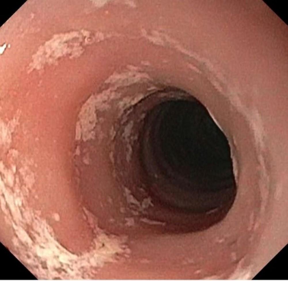
Image provided by Kristle Lynch, MD.
A barium swallow may show stacked circular rings, longitudinal furrows, a narrow-caliber esophagus, or strictures.
Impedance planimetry is occasionally used in patients with significant symptoms to evaluate for subtle strictures.
Testing for food allergies is often done to identify possible triggers but is of minimal benefit because eosinophilic esophagitis is not thought to be IgE-mediated.
Treatment of Eosinophilic Esophagitis
Proton pump inhibitors
Topical corticosteroids
Dupilumab
Elimination diet
Sometimes esophageal dilation
(See also the American Gastroenterological Association (AGA) and the Joint Task Force on Allergy-Immunology Practice Parameters' (JTF) 2020 clinical guidelines for the management of eosinophilic esophagitis.)
In children, PPIs are typically used if dietary changes are ineffective. PPIs are thought to work via the eotaxin-3 pathway.
budesonide is given for at least 8 weeks to determine efficacy. If the patient achieves remission with either of these therapies, they are often continued indefinitely. Maintenance doses of these medications are not well established.
dupilumab had improved histologic outcomes and symptoms (1).
Elimination diets can be effective for some patients in the management of eosinophilic esophagitis (2). The elemental diet may be successful in both adults and children but is often not practical in adults.
Patients who have significant strictures may need careful esophageal dilation using a balloon or bougie; multiple, careful, progressive dilations are done to help prevent esophageal perforation.
Injection and infusion therapies that target the eosinophil pathway in the body are being studied for eosinophilic esophagitis.

Treatment references
1. Dellon ES, Rothenberg ME, Collins MH, et al: Dupilumab in Adults and Adolescents with Eosinophilic Esophagitis. N Engl J Med 387(25):2317-2330, 2022. doi: 10.1056/NEJMoa2205982
2. Mayerhofer C, Kavallar AM, Aldrian D, et al: Efficacy of Elimination Diets in Eosinophilic Esophagitis: A Systematic Review and Meta-analysis. Clin Gastroenterol Hepatol 21(9):2197-2210.e3, 2023. doi: 10.1016/j.cgh.2023.01.019
More Information
The following English-language resources may be useful. Please note that THE MANUAL is not responsible for the content of these resources.
American College of Gastroenterology: Evidenced Based Approach to the Diagnosis and Management of Esophageal Eosinophilia and Eosinophilic Esophagitis (EoE) (2013)
American Gastroenterological Association and the Joint Task Force on Allergy-Immunology Practice Parameters: Clinical guidelines for the management of eosinophilic esophagitis (2020)


