


Etiology
Acute febrile neutrophilic dermatosis may occur with various disorders. It is often classified into 3 categories:
Classical
Cancer-associated
Drug-induced
Disorders and Drugs Associated with Acute Febrile Neutrophilic Dermatosis
Classification | Disorder/Drug |
|---|---|
Classical | Acute respiratory illness Gastrointestinal infection Inflammatory and autoimmune disorders Pregnancy |
Cancer-associated | |
Drug-induced | Granulocyte colony-stimulating factor (G-CSF, the most common drug cause) Anticancer drugs Antiseizure drugs |
About 25% of patients have an underlying cancer, 75% of which are hematologic cancers, especially myelodysplastic syndromes and acute myelogenous leukemia. The dermatosis often precedes the cancer diagnosis. Classical acute febrile neutrophilic dermatosis affects mostly women ages 30 to 50, with a female:male ratio of 3:1. In contrast, men who develop the condition tend to be older (60 to 90). In children under age 3, the ratio is male:female 2:1.
The cause of acute febrile neutrophilic dermatosis is unknown; however, type 1 helper T-cell cytokines, including interleukin-2 and interferon-gamma, are predominant and may play a role in lesion formation.
Symptoms and Signs
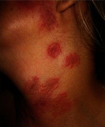
Patients are febrile, with an elevated neutrophil count, and have painful, tender, and edematous red to violet plaques or papules, most often on the face, neck, and upper extremities, especially the dorsum of hands. Oral lesions can also occur. The lesions often develop in crops and may appear annular. Each crop is usually preceded by fever and persists for days to weeks. Rarely, bullous and pustular lesions are present as well.
Less common variants include a bullous form that can ulcerate and resemble pyoderma gangrenosum and a subcutaneous form involving the subcutaneous fat that typically has 2- to 3-cm erythematous nodules, commonly affecting the extremities. When on the lower extremities, this form can resemble erythema nodosum.

Extracutaneous manifestations are rare and can involve the eyes (eg, conjunctivitis, episcleritis, iridocyclitis), joints (eg, arthralgia, myalgia, arthritis), and internal organs (eg, neutrophilic alveolitis; sterile osteomyelitis; psychiatric or neurologic changes; transient kidney, liver, and pancreatic insufficiency).
Diagnosis
Clinical evaluation
Skin biopsy
Diagnosis of acute febrile neutrophilic dermatosis is suggested by the appearance of the lesions and is supported by the presence of associated conditions or drugs. Differential diagnosis can include erythema multiforme, erythema elevation diutinum, subacute cutaneous lupus erythematosus, pyoderma gangrenosum, and erythema nodosum.
If the diagnosis is unclear, skin biopsy should be done. The histopathologic pattern is that of edema in the upper dermis with a dense infiltrate of neutrophils in the dermis. Vasculitis may be present but is secondary.
A complete blood count (CBC) is also done. If the CBC is abnormal, bone marrow biopsy should be considered to diagnose occult cancer.
Treatment
Systemic corticosteroids
Key Points
Acute febrile neutrophilic dermatosis can occur in patients who have certain disorders (classical form) or take certain drugs (drug-induced form), but about 25% of patients have an underlying cancer (cancer-associated form), usually a hematologic cancer.
Diagnose acute febrile neutrophilic dermatosis based on the appearance of the lesions and presence of an associated disorder or drug, and confirm with biopsy when necessary.


