


Edema is swelling of soft tissues due to increased interstitial fluid. The fluid is predominantly water, but protein and cell-rich fluid can accumulate if there is infection or lymphatic obstruction.
Edema may be generalized or local (eg, limited to a single extremity or part of an extremity). It sometimes appears abruptly; patients complain that an extremity suddenly swells. More often, edema develops insidiously, beginning with weight gain, puffy eyes at awakening in the morning, and tight shoes at the end of the day. Slowly developing edema may become massive before patients seek medical care.

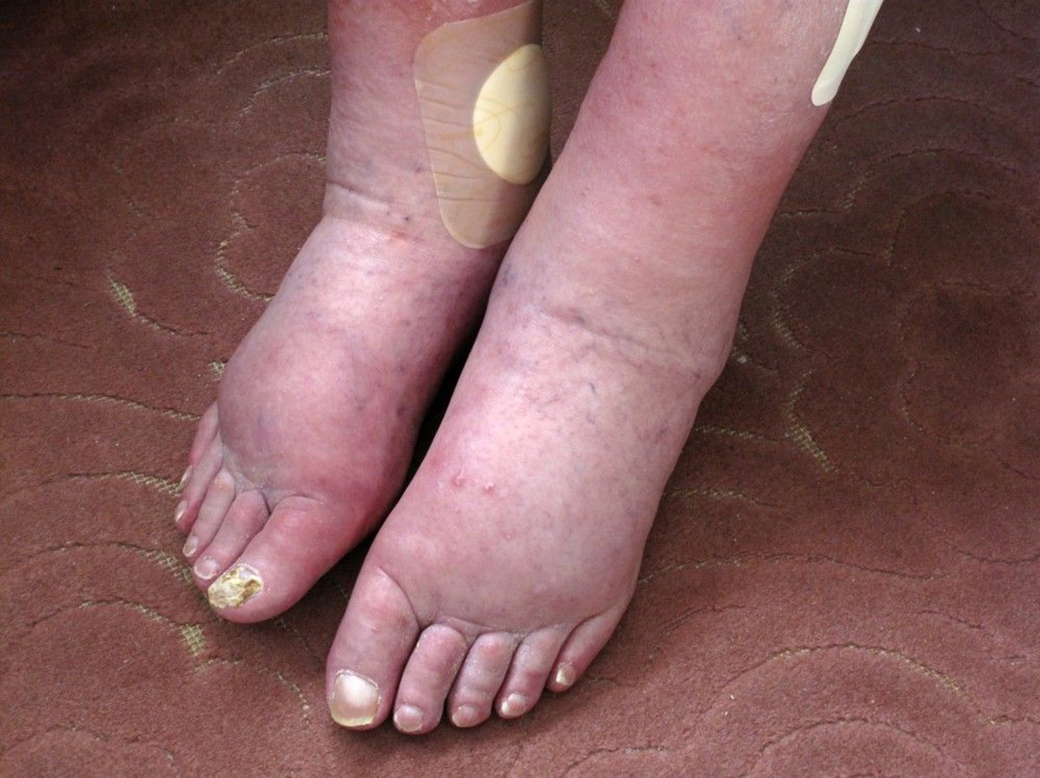
Peter Skinner/SCIENCE PHOTO LIBRARY
Edema itself causes few symptoms other than occasionally a feeling of tightness or fullness; other symptoms are usually related to the underlying disorder. Patients with edema due to heart failure (a common cause) often have dyspnea during exertion, orthopnea, and paroxysmal nocturnal dyspnea. Patients with edema due to deep venous thrombosis (DVT) often have leg pain.


DR P. MARAZZI/SCIENCE PHOTO LIBRARY
Edema due to extracellular fluid volume expansion is often dependent. Thus, in ambulatory patients, edema is in the feet and lower legs; patients requiring bed rest develop edema in the buttocks, genitals, and posterior thighs. Women who lie on only one side may develop edema in the dependent breast. Lymphatic obstruction causes edema distal to the site of obstruction.
Pathophysiology of Edema
Edema results from increased movement of fluid from the intravascular to the interstitial space or decreased movement of water from the interstitium into the capillaries or lymphatic vessels. The mechanism involves one or more of the following:
Increased capillary hydrostatic pressure
Decreased plasma oncotic pressure
Increased capillary permeability
Obstruction of the lymphatic system
As fluid shifts into the interstitial space, intravascular volume is depleted. Intravascular volume depletion activates the renin-angiotensin-aldosterone-vasopressin (ADH) system, resulting in renal sodium retention. By increasing osmolality, renal sodium retention triggers water retention by the kidneys and helps maintain plasma volume. Increased renal sodium retention also may be a primary cause of fluid overload and hence edema. Excessive exogenous sodium intake may also contribute.
Less often, edema results from decreased movement of fluid out of the interstitial space into the capillaries due to lack of adequate plasma oncotic pressure as in nephrotic syndrome, protein-losing enteropathy, liver failure, or starvation.
Increased capilliary permeability occurs in infections or as the result of toxin or inflammatory damage to the capillary walls. In angioedema, mediators, including mast cell–derived mediators (eg, histamine, leukotrienes, prostaglandins) and bradykinin and complement-derived mediators, cause focal edema.
The lymphatic system is responsible for removing protein and white blood cells (along with some water) from the interstitium. Lymphatic obstruction allows these substances to accumulate in the interstitium.
Etiology of Edema
Generalized edema is most commonly caused by
Kidney disorders (especially nephrotic syndrome)
Localized edema is most commonly caused by
DVT or another venous disorder or venous obstruction (eg, by tumor)
Infection
Chronic venous insufficiency may involve one or both legs.
Common causes are listed by primary mechanism (see table Some Causes of Edema).
Evaluation of Edema
History
History of present illness should include location and duration of edema and presence and degree of pain or discomfort. Female patients should be asked whether they are pregnant and whether edema seems related to menstrual periods. Having patients with chronic edema keep a log of weight gain or loss is valuable.
Review of systems should include symptoms of causative disorders, including dyspnea during exertion, orthopnea, and paroxysmal nocturnal dyspnea (heart failure); alcohol or hepatotoxin exposure, jaundice, and easy bruising (a liver disorder); malaise and anorexia (cancer or a liver or kidney disorder); and immobilization, extremity injury, or recent surgery (DVT).
Past medical history should include any disorders known to cause edema, including heart, liver, and kidney disorders and cancer (including any related surgery or radiation therapy). The history should also include predisposing conditions for these causes, including streptococcal infection, recent viral infection (eg, hepatitis), alcohol use disorder, and hypercoagulable disorders. Drug history should include specific questions about drugs known to cause edema (see table Some Causes of Edema). Patients are asked about the amount of sodium used in cooking and at the table.
Physical examination
The area of edema is identified and examined for extent, warmth, erythema, and tenderness; symmetry or lack of it is noted. Presence and degree of pitting (visible and palpable depressions caused by pressure from the examiner’s fingers on the edematous area, which displaces the interstitial fluid) are noted.
In the general examination, the skin is inspected for jaundice, bruising, and spider angiomas (suggesting a liver disorder).
Lungs are examined for dullness to percussion, reduced or exaggerated breath sounds, crackles, rhonchi, and a pleural friction rub.
The internal jugular vein height, waveform, and reflux are noted.
The heart is palpated for thrills, thrust, parasternal lift, and asynchronous abnormal systolic bulge. Auscultation for loud pulmonic component of 2nd heart sound (P2), 3rd (S3) or 4th (S4) heart sounds, murmurs, and pericardial rub or knock is done; all suggest cardiac origin.
The abdomen is inspected, palpated, and percussed for ascites, hepatomegaly, and splenomegaly to check for a liver disorder or heart failure. The kidneys are palpated, and the bladder is percussed. An abnormal abdominal mass, if present, should be palpated.
Red flags
Certain findings raise suspicion of a more serious etiology of edema:
Sudden onset
Significant pain
Shortness of breath
Fever
History of a heart disorder or an abnormal cardiac examination
Hemoptysis, dyspnea, or pleural friction rub
Hepatomegaly, jaundice, ascites, splenomegaly, or hematemesis
Unilateral leg swelling with tenderness
Interpretation of findings
Potential acute life threats, which typically manifest with sudden onset of focal edema, must be identified. Such a presentation suggests acute DVT, soft-tissue infection, or angioedema. Acute DVT may lead to pulmonary embolism (PE), which can be fatal. Soft-tissue infections range from minor to life threatening, depending on the infecting organism and the patient’s health. Acute angioedema sometimes progresses to involve the airway, with serious consequences.
Dyspnea may occur with edema due to heart failure, DVT if PE has occurred, acute respiratory distress syndrome, or angioedema that involves the airways.
Generalized, slowly developing edema suggests a chronic heart, kidney, or liver disorder. Although these disorders can also be life threatening, complications tend to take much longer to develop.
These factors and other clinical features help suggest the cause (see table Some Causes of Edema).
Testing
liver tests, serum protein, and urinalysis (particularly noting the presence of protein and microscopic hematuria). Other tests should be done based on the suspected cause (see table Some Causes of Edema)—eg, brain natriuretic peptide (BNP) for suspected heart failure or D-dimer for suspected pulmonary embolism.
Patients with isolated lower-extremity swelling should usually have venous obstruction excluded by ultrasonography.
Treatment of Edema
Specific causes are treated.
Patients with sodium retention often benefit from restriction of dietary sodium. Patients with heart failure should eliminate salt in cooking and at the table and avoid prepared foods with added salt.
Patients with advanced cirrhosis or nephrotic syndrome often require more severe sodium restriction (≤hyperkalemia can result.
1–4). They can be used in patients with heart failure or nephrotic syndrome with or without diabetes.
Treatment references
1. Anker SD, Butler J, Filippatos G, et al: Empagliflozin in heart failure with a preserved ejection fraction. N Engl J Med 385(16):1451–1461, 2021. doi: 10.1056/NEJMoa2107038
2. Cowie MR, Fisher M: SGLT2 inhibitors: mechanisms of cardiovascular benefit beyond glycaemic control. Nat Rev Cardiol 17(12):761–772, 2020. doi: 10.1038/s41569-020-0406-8
3. Heerspink HJL, Stefánsson BV, Correa-Rotter R, et al: Dapagliflozin in patients with chronic kidney disease. N Engl J Med 383(15):1436–1446, 2020. doi: 10.1056/NEJMoa2024816
4. Packer M, Anker SD, Butler J, et al: Cardiovascular and renal outcomes with empagliflozin in heart failure. N Engl J Med 383(15):1413–1424, 2020. doi: 10.1056/NEJMoa2022190
Geriatrics Essentials: Edema
In older people, use of drugs that treat causes of edema (particularly heart failure) requires special caution, such as the following:
Starting doses low and evaluating patients thoroughly when the dose is changed
Frequently testing for hypokalemia or hyperkalemia
Not stopping calcium channel blockers because of pedal edema, which is benign
Logging daily weight helps in monitoring clinical improvement or deterioration immensely.
Key Points
Edema may result from a generalized or local process and may occur anywhere in the body.
Main causes of generalized edema are chronic heart, liver, and kidney disorders.
Not all edema is serious; consequences depend mainly on the cause.
Sudden onset should trigger prompt evaluation.


