


Pulmonary edema is acute, severe left ventricular failure with pulmonary venous hypertension and alveolar flooding. Findings are severe dyspnea, diaphoresis, wheezing, and sometimes blood-tinged frothy sputum. Diagnosis is clinical and by chest x-ray. Treatment is with oxygen, IV nitrates, diuretics, and, in patients with heart failure and reduced ejection fraction, sometimes short-term IV positive inotropes and assisted ventilation (ie, endotracheal intubation with mechanical ventilation or bilevel positive airway pressure ventilation).
(See also Heart Failure)
If left ventricular (LV) filling pressure increases suddenly, plasma fluid moves rapidly from pulmonary capillaries into interstitial spaces and alveoli, causing pulmonary edema. Although precipitating causes vary by age and country, about one half of cases result from acute coronary ischemia; some from decompensation of significant underlying heart failure (HF), including HF with preserved ejection fraction (HFpEF) due to hypertension; and the rest from arrhythmia, an acute valvular disorder, or acute volume overload often due to IV fluids. Drug or dietary nonadherence is often involved.
Symptoms and Signs of Pulmonary Edema
Patients with pulmonary edema present with extreme dyspnea, restlessness, and anxiety with a sense of suffocation. Cough, possibly producing blood-tinged sputum, pallor, cyanosis, and marked diaphoresis are common; some patients froth at the mouth. Frank hemoptysis is uncommon. The pulse is rapid and low volume, and blood pressure (BP) is variable. Marked hypertension indicates significant cardiac reserve; hypotension with systolic BP < 100 mg Hg is ominous. Inspiratory fine crackles are widely dispersed anteriorly and posteriorly over both lung fields. Marked wheezing (cardiac asthma) may occur. Noisy respiratory efforts often make cardiac auscultation difficult; a summation gallop—merger of 3rd (S3) and 4th (S4) heart sounds—may be present. Signs of right ventricular (RV) failure (eg, neck vein distention, peripheral edema) may be present.
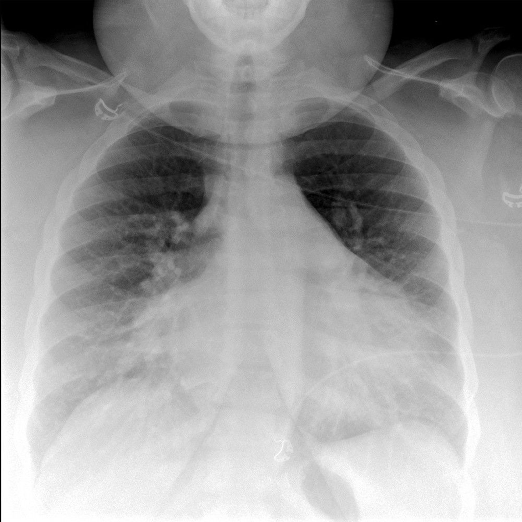
Diagnosis of Pulmonary Edema
Clinical evaluation showing severe dyspnea and pulmonary crackles
Chest x-ray
Sometimes serum brain natriuretic peptide (BNP) or N-terminal-pro BNP (NT-pro-BNP)
ECG, cardiac markers, and other tests for etiology as needed
A COPD (chronic obstructive pulmonary disease) exacerbation can mimic pulmonary edema due to LV failure or even that due to biventricular failure if cor pulmonale is present. Pulmonary edema may be the presenting symptom in patients without a history of cardiac disorders, but COPD patients with such severe symptoms usually have a history of COPD, although they may be too dyspneic to relate it.

© 2017 Elliot K. Fishman, MD.
A chest x-ray, done immediately, is usually diagnostic, showing marked interstitial edema. Bedside measurement of serum BNP/NT-proBNP levels (elevated in pulmonary edema; normal in COPD exacerbation) is helpful if the diagnosis is in doubt.
Echocardiography may be helpful to determine the cause of the pulmonary edema (eg, myocardial infarction, valvular dysfunction, hypertensive heart disease, dilated cardiomyopathy) and may influence the choice of therapies.
Hypoxemia can be severe. Carbon dioxide retention is a late, ominous sign of secondary hypoventilation.
Treatment of Pulmonary Edema
Treatment of cause
Oxygen
IV diuretic
Nitrates
IV inotropes
Ventilatory assistance
>tracheal intubation and mechanical ventilation are required.
Specific additional treatment depends on etiology:
For acute myocardial infarction or another acute coronary syndrome, thrombolysis or direct percutaneous coronary angioplasty with or without stent placement
For severe hypertension, an IV vasodilator
For supraventricular or ventricular tachycardia, direct-current cardioversion
In patients with acute MI (myocardial infarction), fluid status before onset of pulmonary edema is usually normal, so diuretics are less useful than in patients with acute decompensation of chronic heart failure and may precipitate hypotension. If systolic blood pressure falls <
1). Omecamtiv mecarbil, an oral cardiac myosin activator, has been shown to reduce morbidity and mortality in patients currently or recently hospitalized with decompensated heart failure (2).
Once patients are stabilized, long-term HF treatment is begun.
Treatment references
1. Metra M, Teerlink JR, Cotter G, et al: Effects of serelaxin in patients with acute heart failure. N Engl J Med 381(8):716-726, 2019. doi:10.1056/NEJMoa1801291
2. Teerlink JR, Diaz R, Felker GM, et al: Cardiac myosin activation with omecamtiv mecarbil in systolic heart failure. N Engl J Med 384(2):105-116, 2021. doi:10.1056/NEJMoa2025797
Key Points
Acute pulmonary edema can result from acute coronary ischemia, decompensation of underlying heart failure, arrhythmia, an acute valvular disorder, or acute volume overload.
Patients have severe dyspnea, diaphoresis, wheezing, and sometimes blood-tinged frothy sputum.
Clinical examination and chest x-ray are usually sufficient for diagnosis; ECG, cardiac markers, and sometimes echocardiography are done to identify cause.


