


X-rays are a type of medical imaging that use very low-dose radiation waves to take pictures of bones and soft tissues.
X-rays may be used alone (conventional x-ray imaging) or combined with other techniques, such as computed tomography (CT). (See also Overview of Imaging Tests and Background radiation.)
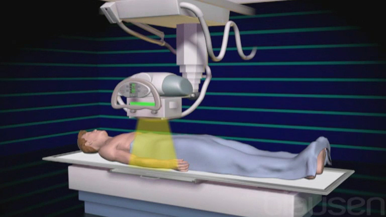
Procedure for X-Rays
For conventional x-ray imaging, a person is positioned so that the body part to be evaluated is between the x-ray source and a device that records the image. The examiner goes behind a screen that blocks x-rays and runs the x-ray machine for only a fraction of a second. The person must remain still when the x-ray is taken. Several x-rays may be taken to obtain images from different angles.
An x-ray beam is aimed at the body part to be evaluated. Different tissues block different amounts of the x-rays, depending on the tissue’s density. The x-rays that pass through are recorded on a film or radiation detector plate, producing an image that shows the different levels of tissue density. The denser the tissue, the more x-rays it blocks and the whiter the image:
Metal appears completely white (radiopaque).
Bone appears almost white.
Fat, muscle, and fluids appear as shades of gray.
Air and gas appear black (radiolucent).
Uses of X-Rays
X-rays are typically the first imaging test done to evaluate the arms, legs, or chest and sometimes the spine and abdomen. These body parts contain important structures with very different densities that are easily distinguished on x-rays. Thus, x-rays are used to detect the following:
Fractures: The almost white bone contrasts clearly with the gray muscles around it.
Pneumonia: The black air in the lungs contrasts clearly with the white infected tissues, which block more of the x-rays.
Blockages of the intestine: The black air in the blocked intestine contrasts clearly with the gray surrounding tissues.
Mammography
In mammography, x-rays are used to screen and diagnose breast disorders, including breast cancer.
Radiation exposure is a concern because breast tissue is sensitive to radiation. Specialized mammography units and digital imaging techniques are used to minimize radiation exposure.
Variations of X-Rays
X-rays with a radiopaque contrast agent
X-rays can be done after a radiopaque contrast agent (sometimes inaccurately called dye) is given, usually by injection into a vein, by mouth, or injected through a tube into the rectum. The radiopaque contrast agent makes the tissue or structure being imaged appear more radiopaque (whiter) than surrounding tissues, so that it can be better seen on an x-ray.
In conventional angiography, x-rays are taken after a radiopaque contrast agent is injected into blood vessels.
Before x-rays of the gastrointestinal tract, people may be asked to swallow barium or gastrografin (which are radiopaque contrast agents) in a liquid or food. The x-rays then show the esophagus, stomach, and small intestine outlined by the barium or gastrografin. Or, an examiner may inject barium through a tube inserted into the anus (barium enema), then carefully pump air into the lower part of the intestine (colon) to expand it. Barium makes ulcers, tumors, blockages, polyps, and diverticulitis easier to detect. A barium enema may cause mild to moderate crampy pain and an urge to defecate.
For imaging of the esophagus, stomach, and upper intestinal tract, endoscopy has largely replaced x-rays taken after barium or gastrografin is used.
Fluoroscopy
Fluoroscopy uses continuous x-ray images to show motion, similar to those of a video camera. Fluoroscopy can show organs or structures as they function: the heart beating, the intestines moving food along, or the lungs inflating and deflating.
Fluoroscopy is commonly used
During electrophysiologic testing (for abnormal heart rhythms) and during coronary catheterization to determine whether a catheter is correctly placed in the heart
With a radiopaque contrast agent (such as barium), usually given by mouth, to evaluate the gastrointestinal tract
During evaluation of musculoskeletal injuries to observe movement of bones and joints
Disadvantages of X-Rays
Other imaging tests may provide better detail, be safer or faster, or help doctors diagnose a disorder more accurately than conventional x-rays.
The main disadvantage of x-rays is
Exposure to radiation
Radiation exposure
For conventional x-rays, each image requires only a very small amount of radiation. For chest x-rays, the amount of radiation exposure with a single image is similar to the amount most people get from the environment in 2.4 days (background radiation exposure).
However, some x-ray tests require several images, a higher dose of radiation for each image, or both. As a result, the total radiation exposure is higher, as in the following examples:
For x-rays of the lower back, done in a series: The amount of radiation equals about 3 months of background exposure.
For mammography, the amount equals about 1 to 2 months of background exposure.
Fluoroscopy usually requires higher doses of radiation than routine x-rays, so other imaging tests are done instead when possible.
Examiners take precautions to minimize a person’s exposure to radiation. Women who are or could be pregnant should tell their doctor. Then, the examiner can take all possible precautions to shield the fetus from exposure. To evaluate the abdomen or pelvis of a pregnant woman, the doctor can sometimes substitute an imaging test that does not use radiation, such as ultrasonography. However, conventional x-rays that do not involve the abdomen or pelvis usually expose the uterus to only very small amounts of radiation.
Other disadvantages
Some particular types of x-rays have other risks. For example, barium swallowed or inserted by enema may cause constipation.


