


A hyphema is bleeding into the front chamber (the fluid-filled space between the clear cornea and the colored iris) of the eye. Additional bleeding (rebleeding) may occur up to several days after the injury.
(See also Overview of Eye Injuries.)
A hyphema will usually resolve with medical treatment, but requires monitoring as it can result in permanent, partial, or complete loss of vision. Vision loss may be caused by increased pressure within the eye (glaucoma), by blood staining the cornea, or both.
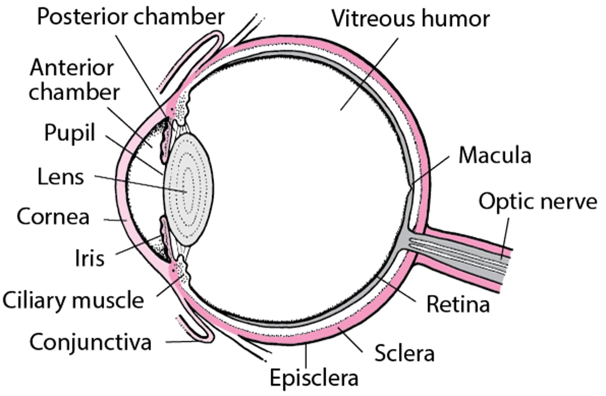
An Inside Look at the Eye


© Springer Science+Business Media
People with hyphema often have blurred vision and pain when exposed to bright light. If the hyphema is large enough, a layer of blood is visible behind the lower part of the cornea when the person is upright. However, the layer may be so small that it can be seen only with magnification.
Treatment of Hyphema
Bed rest with the head of the bed elevated
A protective shield over the eye
Eye drops
A person with a hyphema should be examined by an ophthalmologist (a medical doctor who specializes in the evaluation and treatment—surgical and nonsurgical—of eye disorders) as soon as possible. Some people with severe bleeding or bleeding disorders (which make bleeding and rebleeding more likely) or who take anticoagulant drugs may need to be treated in the hospital.
Doctors measure pressure in the eye at least once daily for the first few days. This measurement is taken painlessly with an instrument called a tonometer


