


In vasa previa, membranes that contain blood vessels connecting the umbilical cord and placenta lie across or near the opening of the cervix—the entrance to the birth canal.
Vasa previa may cause massive bleeding in the fetus and mother when the membranes around the fetus rupture, usually just before labor starts.
To confirm the diagnosis, doctors insert an ultrasound device into the vagina to check for blood vessels over or near the opening of the cervix.
If a woman has vasa previa, doctors check the fetus's heart rate frequently after 28 weeks of pregnancy to determine whether the fetus is in distress.
Cesarean delivery is required and is often done at 34 to 37 weeks or, if problems develop, even earlier.
Vasa previa is present in about 1 in 2,500 to 5,000 deliveries. It is more likely to occur when certain other abnormalities in the placenta are present.
Normally, blood vessels between the fetus and placenta are contained in the umbilical cord. In vasa previa, some of these blood vessels are located in the membranes that surround the fetus, in the area between the fetus and the opening of the cervix. When the membranes rupture, usually a little before labor starts, these blood vessels can be torn. As a result, the fetus may lose a substantial amount of blood. If bleeding is severe, the fetus may die, and the mother may have complications due to massive blood loss.
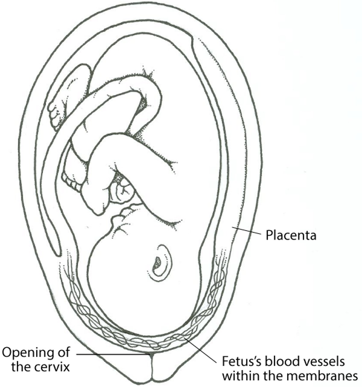
What Is Vasa Previa?
In vasa previa, membranes that contain blood vessels from the fetus to the placenta cross the entrance to the birth canal (the opening of the cervix). When the membranes rupture (near the start of labor), these blood vessels can be torn. |

Symptoms of Vasa Previa
Typically, women have painless vaginal bleeding when the membranes rupture, usually soon after labor starts. The fetus's heart rate is often slow.
Diagnosis of Vasa Previa
Ultrasonography
Doctors may suspect vasa previa when ultrasonography, routinely done earlier in the pregnancy, detects certain abnormalities in the placenta or when the fetus's heart rate is abnormal. Ultrasonography, usually done with a device inserted into the vagina (called transvaginal ultrasonography), can show the blood vessels crossing over or near the opening of the cervix and thus confirm the diagnosis.
Treatment of Vasa Previa
Monitoring of the fetus
Cesarean delivery
If vasa previa is diagnosed before delivery, doctors usually do nonstress testing once or twice a week beginning at 28 to 30 weeks to check on the well-being of the fetus. Doctors may suggest hospitalizing the woman at about 30 to 34 weeks of pregnancy or 1 to 2 weeks before scheduled delivery to closely monitor the fetus.
Women are usually given a corticosteroid to help the fetus's lungs mature.
If vasa previa does not cause any complications, doctors often plan to deliver the baby between 34 to 37 weeks of pregnancy. However, delivery can be earlier if the woman or fetus is in danger.
Delivery is always cesarean.
An emergency cesarean delivery is usually necessary if
Vaginal bleeding continues.
The membranes have ruptured.
The fetus or mother is in danger.


