


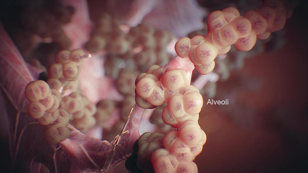
Interstitial lung disease (also called diffuse parenchymal disease) is a term used to describe a number of different disorders that affect the interstitial space of the lungs. The interstitial space consists of the walls of the air sacs of the lungs (alveoli) and the spaces around blood vessels and small airways. Interstitial lung diseases result in abnormal accumulation of inflammatory cells in lung tissue, cause shortness of breath and cough, and have similarities in their appearance on imaging studies but are otherwise unrelated. Some of these diseases are very unusual.
Early in the course of these diseases, white blood cells, macrophages, and protein-rich fluid accumulate in the interstitial space, causing inflammation. If the inflammation persists, scarring (fibrosis) may replace normal lung tissue. As alveoli are progressively destroyed, thick-walled cysts (called honeycombing because they resemble the cells of a beehive) are left in their place. The condition resulting from these changes is called pulmonary fibrosis.
Although the various interstitial lung diseases are separate and have different causes, they have some similar features. All lead to a decreased ability to transfer oxygen to the blood, and all cause stiffening and shrinkage of the lungs, which makes breathing difficult and causes cough. However, being able to eliminate carbon dioxide from the blood is usually not a problem.
Diagnosis of Interstitial Lung Disease
Chest computed tomography
Pulmonary function testing
Arterial blood gas analysis
Because interstitial lung diseases cause symptoms that are similar to those of much more common disorders (for example, pneumonia, chronic obstructive pulmonary disease [COPD]), they may not be suspected at first. When an interstitial lung disease is suspected, diagnostic testing is done. Testing can vary by the disease suspected but tends to be similar.
Most people have a chest x-ray, computed tomography (CT) of the chest, pulmonary function tests, and sometimes arterial blood gas analysis. CT is more sensitive than chest x-ray and helps doctors make a more specific diagnosis. CT is done using techniques that maximize resolution (high-resolution CT). Pulmonary function tests often show that the volume of air that the lungs can hold is abnormally small. Arterial blood gas tests measure the levels of oxygen and carbon dioxide in the arterial blood and determine the acidity (pH) of the blood.
To confirm the diagnosis, doctors sometimes remove a small sample of lung tissue for microscopic examination (lung biopsy) using a procedure called fiberoptic bronchoscopy. A lung biopsy done this way is called transbronchial lung biopsy. Many times, a larger tissue sample is needed and must be removed surgically, sometimes with use of a thoracoscope (a procedure called video-assisted thoracoscopic lung biopsy).
Blood tests may be done. They usually cannot confirm the diagnosis but are done as part of the search for other, similar disorders.


