


Urinary tract obstruction is a blockage that inhibits the flow of urine through its normal path (the urinary tract), including the kidneys, ureters, bladder, and urethra.
Blockage can be complete or partial.
Blockage can lead to kidney damage, kidney stones, and infection.
Symptoms can include pain in the side, decreased or increased urine flow, and urinating at night.
Symptoms are more common if the blockage is sudden and complete.
Testing can include insertion of a urethral catheter, insertion of a viewing tube into the urethra, and imaging tests.
Treatment can include measures to open up a blocked path and to treat the cause of the blockage.
A blockage (obstruction) anywhere along the urinary tract—from the kidneys, where urine is produced, to the urethra, through which urine leaves the body—can increase pressure inside the urinary tract and slow the flow of urine. An obstruction may occur suddenly or develop slowly over days, weeks, or even months. An obstruction may completely or only partially block part of the urinary tract. Sometimes only one kidney is affected, but obstruction may affect both kidneys.
The prevalence of urinary tract obstruction ranges from five in 10,000 to five in 1,000 depending on the cause. In children, obstruction is due mainly to birth defects affecting the urinary tract. Men, particularly those older than 60, are also more likely to be affected because, as men age, the prostate gland tends to increase in size (a condition called benign prostatic hyperplasia) and block the flow of urine.
Hydronephrosis: A Distended Kidney
In hydronephrosis, the kidney is distended because the flow of urine is obstructed. Urine backs up behind the obstruction and remains in the kidney’s small tubes and central collecting area (renal pelvis). |

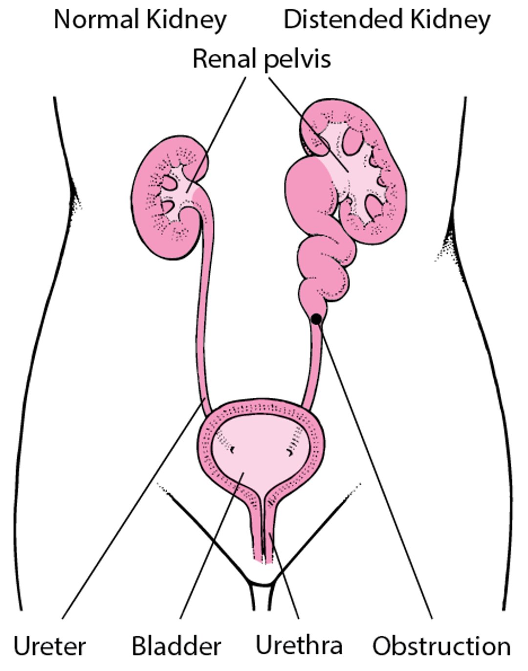
Normally, urine flows out of the kidneys at extremely low pressure. If the flow of urine is obstructed, urine backs up behind the point of blockage, eventually reaching the small tubes of the kidney and its collecting area (renal pelvis), swelling (distending) the kidney and increasing the pressure on its internal structures. Such kidney distention is called hydronephrosis. The elevated pressure due to the obstruction may ultimately damage the kidney and can result in loss of its function.
When the flow of urine is obstructed, stones (calculi) are more likely to form. An infection may develop when the flow of urine is obstructed because bacteria that enter the urinary tract are not flushed out. If both kidneys are obstructed, kidney failure may result.
Long-standing distention of the renal pelvis and ureter can also inhibit the rhythmic muscular contractions that normally move urine down the ureter from the kidney to the bladder (peristalsis). Scar tissue may then replace the normal muscular tissue in the walls of the ureter, resulting in permanent damage.
Partial and complete obstruction tend to cause similar problems, but most problems, and particularly kidney damage, are more severe when obstruction is complete.
Causes of Urinary Obstruction
Blockage may be partial or complete, affect one side or both sides, and develop rapidly (acutely) or slowly (chronically). The most common causes overall are
In children: Structural abnormalities—for example, birth defects such as valves in the inside back part of the urethra (called posterior urethral valves—see Urethra Defects) and other constrictions that narrow or block the ureter or urethra
In young adults: Stones in the kidney or ureter or elsewhere in the urinary tract
In older adults: Benign prostatic hyperplasia (BPH) or prostate cancer, tumors, and stones
Because BPH is so common in older men, obstruction is more common among men. Other common causes of obstruction include strictures (narrowing caused by scar tissue) of the ureter or urethra that develop after radiation therapy, surgery, or procedures done on the urinary tract.
The many other possible causes of urinary tract obstruction include the following:
Polyps in the ureter
Blood clot in the ureter
Tumors in or near the ureter
Disorders of the muscles or nerves in the ureter or bladder (such as due to drugs that have anticholinergic effects [see Anticholinergic: What Does It Mean?], birth defects, or spinal cord injury)
Formation of fibrous (scar) tissue in or around the ureter resulting from surgery, radiation therapy, or drugs (especially methysergide)
Bulging of the lower end of the ureter into the bladder (ureterocele)
Tumor, abscesses, and cysts of the bladder, cervix, uterus, prostate, or other pelvic organs
A large mass of feces stuck in the rectum (rectal impaction)
Hydronephrosis of both kidneys can occur during pregnancy as the enlarging uterus compresses the ureters. Hormonal changes during pregnancy may worsen the problem by reducing the muscular contractions that normally move urine down the ureters. This condition, commonly called hydronephrosis of pregnancy, usually resolves when the pregnancy ends, although the renal pelvis and ureters may remain somewhat distended afterward.
Symptoms of Urinary Obstruction
Symptoms depend on the cause, location, and duration of the obstruction. When the obstruction begins quickly and distends the bladder, ureter, and/or the kidney, it usually causes pain. If the kidney is distended, renal colic can develop. Renal colic is an excruciating pain between the ribs and hip on the affected side that comes and goes every few minutes. The pain may extend into a testis or the vaginal area. People may have nausea and vomiting.
Obstruction of one ureter does not reduce how much people urinate. Obstruction can stop or reduce urination if blockage affects the ureters from both kidneys or if it affects the urethra. Obstruction of the urethra or bladder outlet may cause pain, pressure, and distention of the bladder.
People who have slowly progressive obstruction that causes hydronephrosis may have no symptoms, or they may have attacks of dull, aching discomfort in the flank (the part of the back between the lower end of the ribs and the spine) on the affected side. Sometimes, a kidney stone temporarily blocks the ureter and causes pain that occurs intermittently.
Obstruction that leads to hydronephrosis may cause vague digestive tract symptoms, such as nausea, vomiting, and abdominal pain. These symptoms sometimes occur in children when hydronephrosis results from a birth defect in which the junction of the ureter and renal pelvis is too narrow (ureteropelvic junction obstruction).
People who have urinary tract infections (UTIs) may have pus or blood in the urine, fever, and discomfort in the area of the bladder or kidneys.
Diagnosis of Urinary Obstruction
Bladder catheterization
Imaging
Early diagnosis is important, because most cases of obstruction can be corrected and because a delay in treatment can lead to irreversible kidney damage. Doctors may suspect obstruction because of a person’s symptoms, such as renal colic, symptoms of bladder distention, or a decrease in the volume of urine. A distended kidney can rarely be felt in the flank, usually if the kidney is greatly enlarged in an infant or a child or a thin adult. A distended bladder can sometimes be felt in the lower part of the abdomen just above the pubic bone.
Doctors depend on testing to make the diagnosis.
Bladder catheterization
Bladder catheterization (insertion of a hollow, soft tube through the urethra) is often the first diagnostic test done in people with symptoms that suggest the bladder is distended, such as pelvic pressure or distention. If the catheter drains a large amount of urine from the bladder, then either the bladder outlet or the urethra is obstructed. Many doctors do ultrasonography to determine whether the bladder is filled with a large amount of urine before doing bladder catheterization.
Imaging tests
Imaging tests can be done to identify evidence of obstruction, such as hydronephrosis or a site of blockage, when the presence or site of obstruction is in doubt. For example, ultrasonography is a very useful test in most people (particularly children and pregnant women) because it is fairly accurate and does not expose the person to any radiation. However, ultrasonography is not always accurate in its ability to localize the site of obstruction.
Computed tomography (CT) is an alternative. It is rapid and highly accurate, particularly at identifying stones. CT has traditionally involved exposure to significant doses of radiation. However, with newer CT scanners and new ways of using them, CT images can be obtained with much smaller doses of radiation. Magnetic resonance imaging (MRI) is not as accurate as ultrasonography or CT, particularly for detecting kidney stones, but MRI may be used if it is important to avoid exposing the person to radiation and if the site of obstruction cannot be seen with ultrasonography.
Other imaging tests, such as voiding cystourethrography (VCUG), may be done to identify the site of obstruction, most often in children who have obstruction of the bladder or urethra. This imaging test can identify blockages in those structures (for example, caused by birth defects). It can also identify when urine flows backward from the bladder into the ureters (called vesicoureteral reflux) and causes urinary tract infections (UTIs) as well as obstruction. In VCUG, x-rays are taken after a radiopaque agent (dye) is inserted through a catheter inserted into the bladder.
Endoscopy
Endoscopy with a special rigid or flexible endoscope (a cystoscope) can be used to examine the urethra, prostate, and bladder. A longer rigid or flexible endoscope (ureteroscope) can be passed into the ureters or kidneys to identify sites of obstruction. Sometimes the cystoscope, ureteroscope, or both can also be used to remove objects causing obstruction.
Blood and urine tests
Prognosis for Urinary Obstruction
Blockage can usually be relieved, but if relief takes too long, the kidney can be damaged permanently. However, because one normally functioning kidney is enough to sustain the body, permanent kidney failure is unlikely to develop unless both kidneys have been blocked for some time, usually at least a few weeks. The prognosis also depends on the cause of obstruction. For example, an untreated infection is more likely to cause kidney damage than a kidney stone.
Treatment of Urinary Obstruction
Relief of obstruction
Treatment usually aims to relieve the cause of obstruction. For example, if the urethra is blocked because of a benign enlarged or cancerous prostate, treatment can include drugs, such as hormonal therapy for prostate cancer, surgery, or enlargement of the urethra with dilators. Other treatments, such as lithotripsy or endoscopic surgery, may be needed to remove stones that block the flow of urine in the ureter or kidney.
If the cause of obstruction cannot be rapidly corrected, particularly if there is infection, acute kidney failure, or severe pain, the urinary tract is drained. When acute hydronephrosis is caused by an obstruction that is not easily relieved, urine that has accumulated above the obstruction can be drained with a soft tube inserted through the back into the kidney (nephrostomy tube) or by insertion of a soft plastic tube that connects the bladder with the kidney (ureteral stent). Complications of nephrostomy tubes or ureteral stents can include displacement of the tube, infection, and discomfort. If the urethra is the site of an obstruction that must be relieved rapidly, doctors insert a soft rubber catheter into the bladder to drain urine.
Obstructions that cause chronic hydronephrosis usually do not require urgent relief. Complications of urinary tract obstruction, such as urinary tract infections and acute kidney failure, if present, are treated promptly.


