


Computed tomography (CT) is a type of medical imaging that combines a series of x-rays to create cross-sectional, detailed images of internal structures.
In computed tomography (CT), which used to be called computed axial tomography (CAT), an x-ray source and x-ray detector rotate around a person. In modern scanners, the x-ray detector usually has 4 to 64 or more rows of sensors that record the x-rays that pass through the body. Data from the sensors represent a series of x-ray measurements taken from multiple angles all around the person. However, the measurements are not viewed directly but are sent to a computer. The computer converts them into images that resemble 2-dimensional slices (cross-sections) of the body. The computer can also construct 3-dimensional images from the recorded images.
(See also Overview of Imaging Tests.)

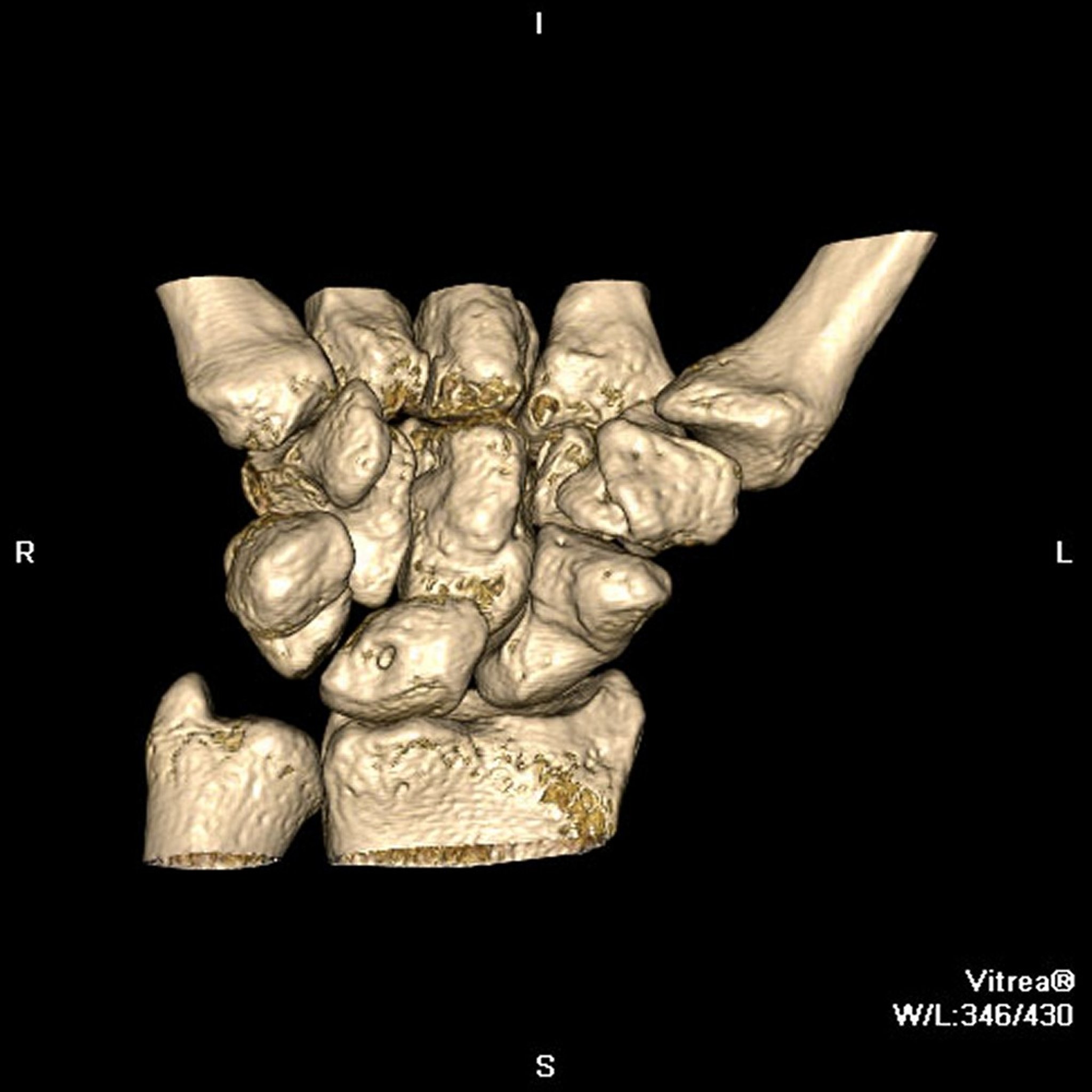
Image provided by Jon A. Jacobson, MD.
Procedure for CT
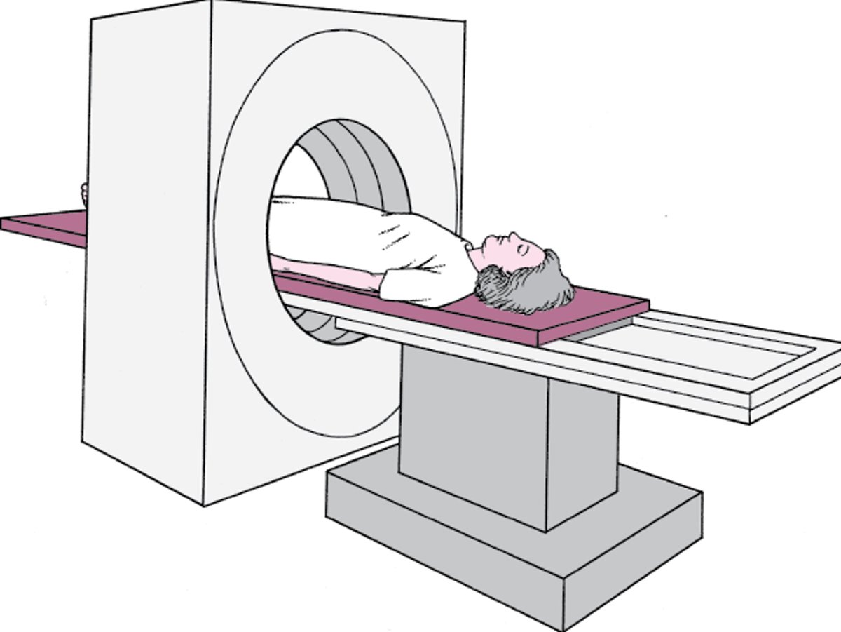
For CT, a person lies on a motorized table that is moved through the opening of a doughnut-shaped scanner. The person is moved through the scanner as these devices rotate around the person. For some CT scans, the table moves incrementally and stops when each scan (slice) is taken. For other CT scans, the table moves continuously during scanning. Because the person is moving in a straight line and the detectors are moving in a circle, the series of measurements appear to be taken in a spiral fashion around the person.
People should wear clothing that has no metallic buttons, snaps, zippers, or other metal in it over the area to be scanned and should remove any jewelry. Such items are not dangerous but may block x-rays and distort the image. During the test, people must remain still and periodically hold their breath when the x-rays are taken so that the images are not blurred. People may hear whirring sounds during the procedure.
The procedure, depending on the area examined and how modern the scanner is, usually takes from only a few seconds to a few minutes. CT of the chest takes less than a minute, and people have to hold their breath only once and only for a few seconds.
For CT, people may be given a radiopaque contrast agent. Contrast agents are substances that can be seen on x-rays and help distinguish one tissue from another. The contrast agent may be injected into a vein, taken by mouth, or inserted through the anus. The contrast agents used depends on what type of test is done and which body part is being evaluated.
CT is usually done as an outpatient procedure. People can resume their usual activities immediately after the test.
Imaging the Interior: Computed Tomography
In computed tomography, a scanner produces and records x-rays as it rotates around a person, who is moved through the scanner on a motorized table. On one side of the scanner is an x-ray tube, which produces x-rays, and on the other side is an x-ray detector. |

Uses of CT
The highly detailed images provide more detail about tissue density and location of abnormalities than x-rays, so doctors can precisely locate structures and abnormalities. CT enables the examiner to distinguish between different types of tissue, such as muscle, fat, and connective tissues. Thus, CT can provide detailed images of specific organs not visible on x-rays and is more useful for imaging most structures in the brain, head, neck, chest, and abdomen.
CT can detect and provide information about disorders in almost every part of the body. For example, doctors can use CT to detect a tumor, measure its size, precisely locate it, and determine how far it has spread into nearby tissues. CT can also help doctors monitor the effectiveness of treatment (such as antibiotics for a brain abscess or radiation therapy for a tumor).
Variations of CT
CT angiography
CT angiography uses CT and a radiopaque contrast agent to produce 2- and 3-dimensional images of blood vessels, including the arteries that supply the heart (coronary arteries). The contrast agent is injected into a vein (not an artery as in conventional angiography), usually in the arm. Images are taken rapidly and are timed so that they show the radiopaque contrast agent flowing through the blood vessels being evaluated. The computer digitally removes all tissues except blood vessels from the images. (See also Coronary Angiography.)
CT angiography is used to detect the following:
Narrowing or blockages (such as blood clots) in arteries
Bulges (aneurysms) and tears (dissections) in large arteries
Abnormal blood vessels that carry blood to tumors
CT angiography is commonly used instead of conventional angiography because it is safe and less invasive (it does not require insertion of a catheter in an artery, which has slightly more risk than insertion of a catheter into a vein). CT angiography shows abnormalities in blood vessels about as accurately as magnetic resonance angiography, but slightly less accurately than conventional angiography.
CT angiography usually takes only 1 to 2 minutes.
Other variations
CT can be used to provide images of the
Stomach or small intestine (called CT enterography)
Colon (called virtual colonoscopy, or CT colonography)
Kidneys, ureters, and bladder (called CT intravenous urography or pyelography)
Arteries of the lungs (called CT pulmonary angiography)
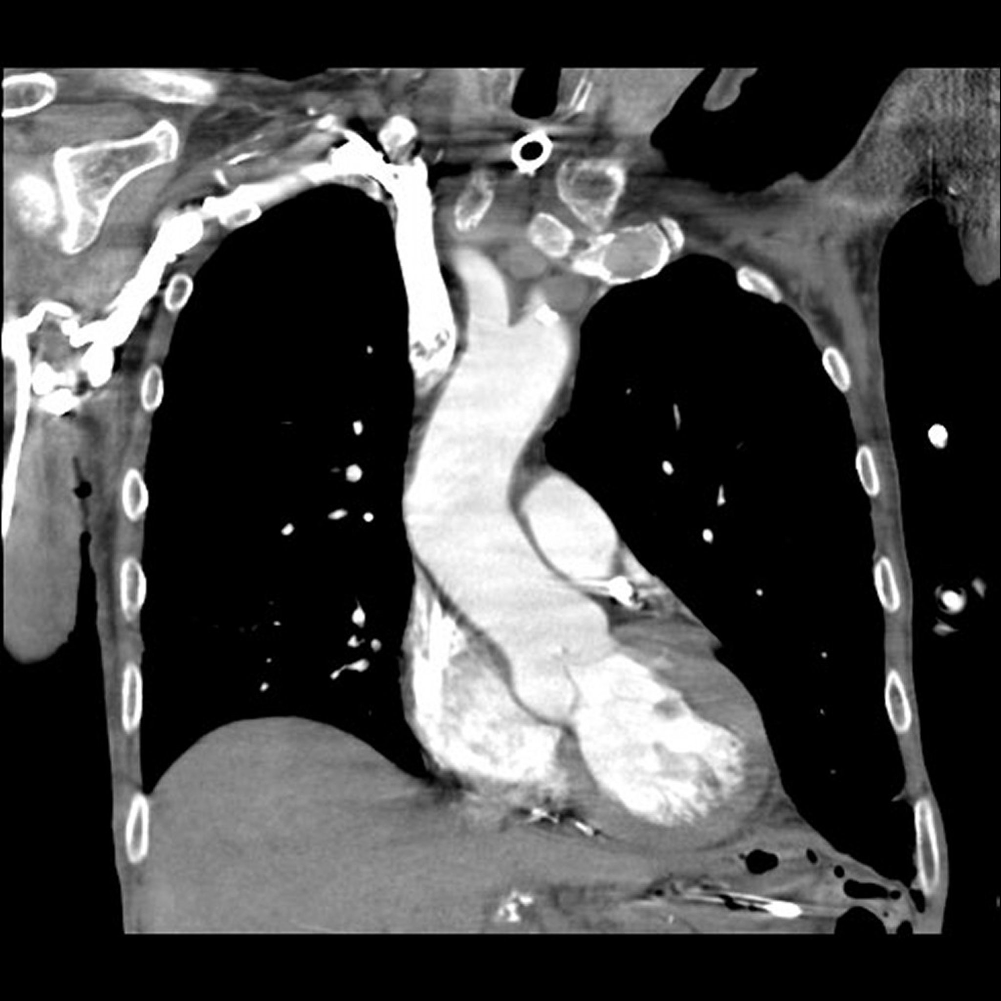
Image provided by Mehmet Kocak, MD.
Disadvantages of CT
Typically, CT of the abdomen uses about 300 to 400 times the amount of radiation used for a single-view x-ray of the chest. Even though newer CT techniques use much lower radiation doses than those previously used, CT accounts for most exposure to man-made radiation in the general population and for about 70% of radiation exposure in medical practice. Therefore, the doctor and person should carefully weigh the benefit of each CT procedure against the risks (see Risks of Radiation). Generally, CT is avoided when possible in pregnant women unless there is no good alternative. Use of CT in children should be limited as much as possible.
The radiopaque contrast agents used in CT angiography contain iodine—called iodinated contrast agents. A few people have a mild to severe allergic reaction or kidney damage after such agents are injected. People who have had reactions to these agents should let their doctor know before CT angiography is done.
In some countries and in some areas of the United States, CT is not readily available.
Did You Know
|
More Information
The following English-language resource may be useful. Please note that THE MANUAL is not responsible for the content of this resource.
What Are the Radiation Risks from CT?: This Food and Drug Administration (FDA) resource explains the various types of imaging tests used for screening and diagnosis and well as the risks and benefits of each.


