


Tinea versicolor is a fungal infection of the topmost layer of the skin that causes scaly, discolored patches.
This infection is caused by a type of fungus.
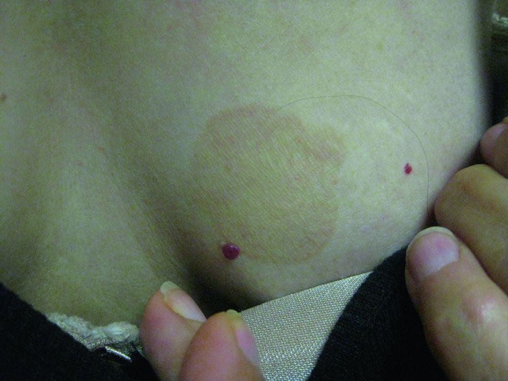
Typically, people have tan, brown, salmon, or white scaly patches of skin.
The diagnosis is based on appearance and skin scrapings.
Antifungal skin products, shampoos, and sometimes medications taken by mouth are used to treat the infection.
Tinea versicolor infection often returns.
(See also Overview of Fungal Skin Infections.)
The infection is caused by Malassezia furfur. Malassezia furfur is a type of fungus that can exist as both a yeast and a mold. Yeast and mold are terms that are used to describe what the fungus looks like under a microscope.
Malassezia furfur is typically harmless and normally lives on the skin but in some people causes tinea versicolor. Most affected people are healthy. Some people may be genetically predisposed to overgrowth of this fungus.
Other risk factors for tinea versicolor include heat and humidity and an immune system weakened by corticosteroid use, pregnancy, undernutrition, diabetes, or other disorders.
Tinea versicolor is a mild infection and is not considered contagious. It is quite common, especially among young adults.
Symptoms of Tinea Versicolor
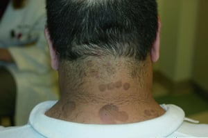
Tinea versicolor causes many tan, brown, salmon, or white scaly patches to appear on the trunk, neck, abdomen, and occasionally the face. The patches may join to form larger patches. The patches do not tan, so in summer, when the surrounding skin tans, the patches may become obvious. People with naturally dark skin may notice lighter patches. People with naturally fair skin may develop darker or lighter patches.
Tinea versicolor usually does not cause other symptoms.
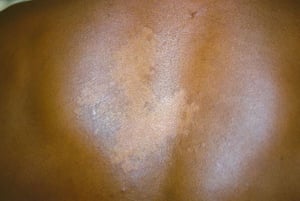
Image courtesy of Karen McKoy, MD.
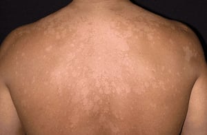
Image provided by Thomas Habif, MD.

© Springer Science+Business Media
© Springer Science+Business Media
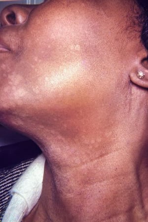
Image courtesy of Karen McKoy, MD.

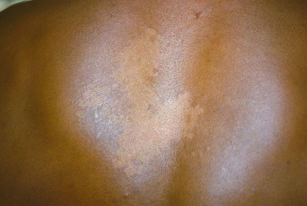
Image courtesy of Karen McKoy, MD.

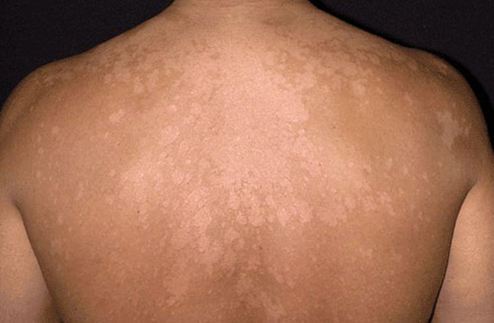
Image provided by Thomas Habif, MD.

© Springer Science+Business Media


© Springer Science+Business Media


Image courtesy of Karen McKoy, MD.
Diagnosis of Tinea Versicolor
A doctor's examination of the skin and skin scrapings
Sometimes a Wood light examination
Doctors diagnose tinea versicolor by the appearance of the skin and by looking at skin scrapings under a microscope to see the fungus/yeast.
Doctors may use an ultraviolet light (called a Wood light) to show the infection on the skin more clearly.
Treatment of Tinea Versicolor
Antifungal medications applied to the affected areas or sometimes taken by mouth
Some Antifungal Medications Applied to the Skin (Topical Medications).)
Medications for Serious Fungal Infections) or frequent infections.
To lower the chance of the infection coming back, many doctors recommend practicing meticulous hygiene and using pyrithione zinc soap regularly or one of the other topical treatments monthly.
Prognosis for Tinea Versicolor
The skin may not regain its normal pigmentation for many months or years after the infection is gone.
Tinea versicolor commonly comes back after successful treatment because the yeast that causes it normally lives on the skin.


