


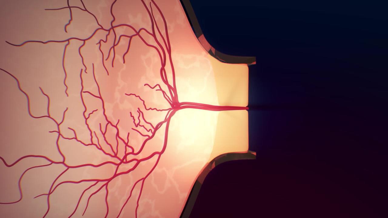
A vein in the retina (the transparent, light-sensitive structure at the back of the eye) may become blocked, causing sudden, painless loss of vision.
Doctors typically make the diagnosis by looking in the eye with an ophthalmoscope and sometimes by diagnostic tests.
Treatment can often improve vision.
The central retinal vein is the main vein that drains blood away from the retina. Blockage may occur in the main vein or in its branches. (See also Overview of Retinal Disorders.)
Central retinal vein blockage occurs mainly in older adults. Risk factors include
Increased blood viscosity (thickness)
Sometimes the cause of the blockage is unknown.
Symptoms of Retinal Vein Blockage
Blockage of the central retinal vein causes severe, painless, and usually sudden loss of vision, but vision loss can also sometimes occur gradually over a period of days to weeks.
Blockage of the central retinal vein may also cause growth of abnormal blood vessels on the retina or iris. Sometimes, these abnormal blood vessels bleed or cause a painful type of glaucoma (called neovascular glaucoma). In neovascular glaucoma, abnormal blood vessels that have formed in the iris close the space between the iris and the cornea, blocking the drainage of fluid from the eye and causing buildup of pressure in the eye (glaucoma). Other complications may include bleeding in the vitreous humor (the clear, gel-like substance in the eyeball that gives it its round shape).
Diagnosis of Retinal Vein Blockage
A doctor's examination of the eye
Optical coherence tomography
Sometimes other tests
Using an ophthalmoscope, doctors can see changes in blood vessels and the retina. If the central retinal vein is blocked, the veins may be engorged (appearing enlarged), bleeding spots may be visible scattered throughout the retina, and the front of the optic nerve may be swollen.
Optical coherence tomography (an imaging study) can help show that the retina is swollen, which is common.
Once retinal vein blockage has been diagnosed, doctors often do tests to identify disorders that could increase the risk of developing blockages. For example, depending on which specific disorders they suspect, doctors may test people for diabetes (by measuring blood sugar or hemoglobin A1C levels), glaucoma (by measuring eye pressure), high blood pressure (by measuring blood pressure), and disorders that cause abnormally thick blood (called hyperviscosity disorders).
Treatment of Retinal Vein Blockage
Medications injected into the eye
Laser treatment of abnormal or bleeding blood vessels
Certain medications can be injected into the eye, or an implant that slowly releases constant levels of a corticosteroid can be injected into the eye. Laser treatment of the leaking blood vessels can also help improve vision for some people with a blockage in a branch of the retinal vein. Although the treatments help restore vision in a number of people, many people have some permanent vision loss. Thus, preventing such blockages by controlling risk factors (for example, high blood pressure, diabetes, and other risk factors for atherosclerosis) is desirable.
Laser treatment may be used to destroy abnormal blood vessels to treat or prevent neovascular glaucoma or prevent further vision loss from bleeding within the eye.
Prognosis for Retinal Vein Blockage
How much vision people with a retinal vein blockage retain depends mainly on 2 things:
Whether the blockage affected the central retinal vein or a branch
Sharpness of vision (visual acuity) at the time of the blockage
Most people have some permanent loss of vision.
If the visual acuity is good at the time of retinal vein blockage (usually when only a branch is blocked), it will likely remain good, occasionally near normal. If the visual acuity is poor (for example, worse than 20/200), it will remain poor or worsen in 80% of people. Blockage of the central retinal vein rarely recurs.
More Information
The following English-language resource may be useful. Please note that THE MANUAL is not responsible for the content of this resource.
National Eye Institute: A resource for learning about eye health (in English and Spanish) for adults and children, as well as for providing access to outreach campaigns.


