


Ischemic optic neuropathy is damage of the optic nerve caused by a blockage of its blood supply.
Blockage can occur with inflammation of the arteries (called arteritic, typically as part of a disorder called giant cell arteritis) or without inflammation of the arteries (called nonarteritic).
The only constant symptom is painless vision loss, which is usually sudden.
Doctors make the diagnosis based on the person's symptoms and by looking in the person’s eye with an ophthalmoscope.
Blood tests and sometimes biopsy of temporal artery tissue are done to diagnose giant cell arteritis.
Treatment for the nonarteritic variety is not effective.
Treatment for the arteritic type does not restore vision but can help protect the unaffected eye.
(See also Overview of Optic Nerve Disorders.)
Causes of Ischemic Optic Neuropathy
Blockage of the blood supply to the part of the optic nerve within the eye can lead to impaired function of optic nerve cells and vision loss. Two types can occur: nonarteritic and arteritic.
Nonarteritic ischemic optic neuropathy occurs more frequently and usually occurs in people about age 50 and older. Vision loss is not usually as severe as in arteritic ischemic optic neuropathy. Risk factors include an anatomically congested optic nerve (small cup-to-disc ratio), high blood pressure, smoking, diabetes, and atherosclerosis. Other risk factors may include obstructive sleep apneaerectile dysfunction), a tendency to develop blood clots, and low blood pressure at night.
Arteritic ischemic optic neuropathy usually occurs in people about age 60 and older. The blood supply to the optic nerve is blocked due to inflammation of the arteries (arteritis), most notably giant cell arteritis.
Symptoms of Ischemic Optic Neuropathy
Loss of vision is usually rapid (over minutes, hours, or rarely days) but is painless. Depending on the cause, vision may be impaired in one or both eyes. Vision in the involved eye or eyes can range from almost normal to complete blindness.
People with giant cell arteritis tend to be older, and their loss of vision tends to be more severe. They may have jaw pain when they chew, headaches, muscle aches and pains, and scalp pain when they comb their hair.
Diagnosis of Ischemic Optic Neuropathy
A doctor's evaluation, including visual field examination
For giant cell arteritis, blood tests and biopsy
Sometimes imaging or other tests
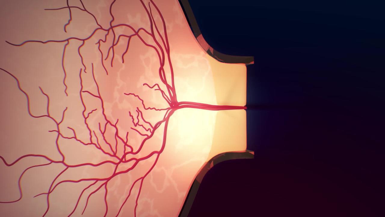
Diagnosis involves examination of the back of the eyes with a light with magnifying lenses (ophthalmoscope) and a visual field examination to measure central or peripheral vision loss. The head of the optic nerve at the back of the eye (optic disc will be swollen. Determining the cause involves determining whether the person has any of the disorders known to be risk factors.
If giant cell arteritis is suspected as a cause, blood tests are done and corticosteroids are started immediately to prevent further vision loss. Removal and examination of a temporal artery tissue sample under a microscope (biopsy) may be done to confirm the diagnosis. Blood tests determine the erythrocyte sedimentation rate (ESR), the C-reactive protein level, and the levels of certain types of blood cells (complete blood count). Results of these tests may indicate inflammation that is characteristic of giant cell arteritis. If a person has no symptoms of giant cell arteritis, magnetic resonance imaging (MRI) or computed tomography (CT) of the brain may be done to make sure the optic nerve is not being compressed by a tumor.
Other tests may be necessary depending on what causes are likely. For example, if people have symptoms of obstructive sleep apnea (such as excessive daytime sleepiness or snoring), polysomnography may be done. If people have had blood clots, blood tests may be done to diagnose blood-clotting disorders.
Prognosis for Ischemic Optic Neuropathy
There is no effective treatment for nonarteritic ischemic optic neuropathy. However, about one third of people with nonarteritic ischemic optic neuropathy have partial improvement of their vision spontaneously. In this condition, repeat episodes in the same eye are rare, but the other eye is affected in 15 to 20% of people.
In the arteritic variety caused by giant cell arteritis, vision loss is typically greater than in nonarteritic ischemic optic neuropathy. Prompt treatment with corticosteroids does not restore lost vision in the affected eye but protects the unaffected eye. Inadequate treatment increases the risk of vision loss in the other eye.
Treatment of Ischemic Optic Neuropathy
For nonarteritic ischemic optic neuropathy, control of risk factors for atherosclerosis
In people with nonarteritic ischemic optic neuropathy, treatment to restore vision loss is ineffective. Treatment involves reducing risk factors for atherosclerosis, including controlling blood pressure and diabetes. Other causes, such as blood-clotting disorders and obstructive sleep apnea, may also require treatment.
In people with arteritic ischemic optic neuropathy caused by giant cell arteritisgiant cell arteritis.
Magnifiers, large-print devices, and talking watches (low-vision aids) may help people with loss of vision.


