


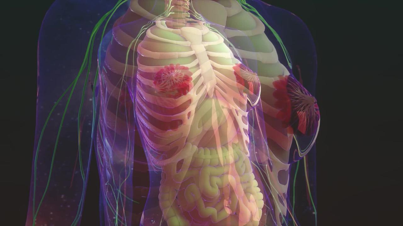
A breast lump (mass) is a thickening or bump that feels different from surrounding breast tissue. A lump may be discovered by a woman or during a breast examination by a doctor.
(See also Overview of Breast Disorders.)
Lumps in the breasts are relatively common and usually not cancerous.
Did You Know...
|
Lumps may be painless or painful. They are sometimes accompanied by a nipple discharge or changes in the skin, such as irregularities, redness, a dimpled texture (called peau d'orange, or skin of an orange), or tightened skin.
Breast lumps may be fluid-filled sacs (cysts) or solid masses, which are usually fibroadenomas. Fibroadenomas are not cancerous, and cysts usually are not cancerous.
Causes of Breast Lumps
Common causes of breast lumps
The most common causes involve the fibroglandular tissue (composed of fibrous connective tissue and glands) in the breast, including
Fibroadenomas
Fibrocystic changes
Fibroadenomas are typically smooth, rounded, movable, painless lumps. They usually develop in women of child-bearing age, and they may decrease in size over time. Fibroadenomas may be mistaken for breast cancer, but they are not. Some types of fibroadenoma do not appear to increase the risk of breast cancer. Others may increase the risk slightly.
Fibrocystic changes includes pain, cysts, and general lumpiness in the breast. Women may have one or more of these symptoms. Breasts feel lumpy and dense and are often tender when touched. These changes are more common among women who began to menstruate early, had their first baby after age 30, or have not had a baby.
In most women, fibrocystic changes are related to the monthly fluctuations in levels of the female hormones estrogen and progesterone. These hormones stimulate breast tissue. Symptoms tend to subside after menopause.
Fibrocystic changes do not increase the risk of breast cancer.
Other causes of breast lumps
Lumps sometimes result from
Breast infections, including collections of pus (abscesses), which are very rare except during the few weeks after childbirth
A clogged milk gland (galactocele), which usually occurs up to 6 to 10 months after breastfeeding stops
Injuries, which can result in the formation of scar tissue
Infections, galactoceles, and scar tissue formation do not increase the risk of breast cancer.
Evaluation of Breast Lumps
Warning signs
Certain symptoms and characteristics are cause for concern:
A lump that is stuck to the skin or chest wall
A lump that is hard and irregular in texture
Dimpling of skin near the lump
Thickened, red skin over the breast
A bloody discharge from the nipple
Lymph nodes in the armpit that are matted together or stuck to the skin or chest wall
When to see a doctor
Because breast lumps may be cancerous (although they usually are not), they should be evaluated by a doctor as soon as possible.
What the doctor does
Doctors ask a woman questions about the lump, such as how long it has been present, whether it comes and goes, and whether it is painful. Doctors also ask about other symptoms, including any discharge from the nipple and general symptoms such as weight loss, fatigue, and bone pain. Doctors ask a woman about her medical and family history, including a previous diagnosis of breast cancer and risk factors for breast cancer.
Doctors then do a breast examination (see Screening). Doctors inspect the breast, looking for abnormalities, changes in the skin, and nipple discharge. They also feel (palpate) the lump to determine
How large it is
Whether it is hard or soft
Whether it is smooth or irregular
Whether it is painful
Whether it moves freely or is stuck to the skin or chest wall
Painful, rubbery lumps in younger women are usually fibrocystic changes, particularly if a woman has had similar lumps before.
Doctors determine whether the breasts are similar in shape and size and check each breast for abnormalities, particularly warning signs. Cancer is more likely if warning signs are present.
Doctors also feel the lymph nodes in the armpits and above the collarbone to check for enlarged or painful lymph nodes.
Testing
If a breast lump is found on breast examination, further testing is needed to determine whether it is cancerous.
Ultrasonography is typically done first to try to differentiate solid lumps from cysts, which are rarely cancerous.
If the lump appears to be a cyst and is causing symptoms (such as pain or nipple discharge), a needle with a syringe may be inserted into the cyst, and the fluid removed (called aspiration) and examined. The fluid is tested for cancer cells only if any of the following occurs:
It is bloody or cloudy.
Little fluid is obtained.
The lump remains after aspiration.
Otherwise, a woman is checked again in 4 to 8 weeks. If the cyst cannot be felt, it is considered noncancerous. If it recurs, aspiration is done again, and the fluid is sent for analysis regardless of appearance. If the cyst recurs a third time or if a lump is still present after it was aspirated, a sample of tissue from the lump or the entire lump is removed and examined under a microscope (biopsy).
If the lump appears to be solid, mammography is typically done, followed by a biopsy. Doctors may do one of several types of biopsy:
Fine-needle aspiration biopsy: Some cells are removed from the lump through a thin needle attached to a syringe.
Core needle biopsy: A larger needle with a special tip is used to remove a larger sample of breast tissue.
Open (surgical) biopsy: Doctors make a small cut in the skin and breast tissue and remove part or all of a lump. This type of biopsy is done when a needle biopsy is not possible (for example, because no lump is felt). It may also be done after a needle biopsy that does not detect cancer to be sure that the needle biopsy did not miss a cancer.
Ultrasonography or mammography is often used to guide placement of the needle for the biopsy. Most women do not need to be hospitalized for these procedures. Usually, only a local anesthetic is needed.
Treatment of Breast Lumps
Treatment of breast lumps and fibrocystic changes depends on what the cause is and whether symptoms are present.
Sometimes cysts are drained.
Fibroadenomas are usually removed if they are enlarging or causing pain or if a woman wants them to be removed. If the fibroadenomas are small, they may be destroyed using cold (cryoablation). Usually for this procedure, only a local anesthetic is required. However, after one fibroadenoma is removed, other fibroadenomas may appear in other parts of the breast. If several lumps have been removed and found to be noncancerous, a woman and her doctor may decide against removing new lumps that develop. Regardless of whether the fibroadenomas are removed or not, a woman should have regular check-ups so that her doctor can check for changes.
If a lump is a galactocele (a clogged milk gland), it is drained (aspirated). It typically resolves after this treatment.
Treatment of breast cancer, if diagnosed, usually consists of surgery to remove the tumor plus radiation therapy, chemotherapy, and/or hormonal medications.
Key Points
Most breast lumps are not cancer.
Women with a breast lump should see a health care professional, who examines the breast and usually does additional tests.


