


Doctors use specific terms to describe various types of marks and growths on the skin. Some skin disorders and infections can cause color changes in the skin.
(See also Structure and Function of the Skin.)
Types of Skin Marks and Growths
Atrophy is thinning of the skin that can sometimes result in a depression and often has a dry and wrinkled "cigarette paper" appearance.
Bullae are fluid-filled blisters that are greater than 10 millimeters (0.4 inch) in diameter (larger than vesicles).
Crusts (scabs) are dried blood, pus, or skin fluids on the surface of the skin. A crust can form wherever the skin has been damaged.
Cysts are thin-walled cavities filled with fluid or semi-liquid material. They often look and feel like a lump (nodule) in the skin.
Erosions are open areas of skin that result from loss of part or all of the top layers (epidermis) of the skin. Erosions occur when infection, pressure, irritation, or temperature has damaged the skin. They typically heal without scarring.
Excoriations are erosions caused by scratching, rubbing, or picking at the skin. Often, excoriations are covered with a crust.
Lesion is a general term for any abnormal mark or growth on the skin.
Lichenification is thickened skin that has accentuated skinfolds or creases that appear as deep grooves and wrinkles. Lichenification is caused by repeated scratching or rubbing.
Macules are flat, discolored spots of any shape less than 10 millimeters (0.4 inch) in diameter. Freckles, flat moles, port-wine stains, and many rashes are macular.
Nodules are solid raised areas that are usually round. They are deeper and easier to feel than papules. A nodule sometimes appears to form below the surface of the skin and press upward.
Papules are raised solid bumps less than 10 millimeters (0.4 inch) in diameter. Warts, insect bites, lichen planus, and some skin cancers can grow as papules.
Patches are larger flat spots (greater than 10 millimeters).
Plaques are flat or raised areas or groups of small bumps (papules) typically more than 10 millimeters (0.4 inch) in diameter.
Pustules are fluid-filled spots (vesicles) containing pus.
Scales are areas of heaped-up, dead epidermal cells that appear as a flaky, dry patch. Scales occur with psoriasis, seborrheic dermatitis, and many other disorders.
Scars are areas where normal skin has been replaced by fibrous (scar-forming) tissue. Scars form after damage of the dermis.
Telangiectases are dilated blood vessels near the surface of the skin that often have a twisted appearance and that whiten (blanch) when pressure is applied.
Ulcers are similar to erosions, only deeper, penetrating at least part of the dermis. The causes are the same as for erosions, but conditions that impair healing, such as venous stasis, diabetes, peripheral artery disease, and vasculitis, are also often involved. Ulcers usually heal with scarring.
Vesicles are small, fluid-filled blisters less than 10 millimeters (0.4 inch) in diameter. Bullae are vesicles larger than 10 millimeters in diameter. Herpes zoster (shingles), chickenpox, burns, allergic reactions, and irritations form vesicles and bullae.
Wheals (hives, urticaria) are elevated, itchy areas that are caused by swelling in the skin. In people who have light skin, hives are usually red. In people who have dark skin, hives may look closer to the color of surrounding skin. Wheals appear relatively suddenly and then almost always disappear within 24 hours. Wheals are common allergic reactions to drugs or medications, insect bites, or something that touches the skin. The presence of multiple wheals is called hives or urticaria.
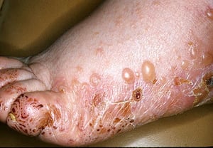
Photo provided by Thomas Habif, MD.

Photo provided by Thomas Habif, MD.
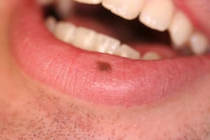
Photo provided by Robert MacNeal, MD.
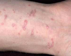
Photo provided by Thomas Habif, MD.
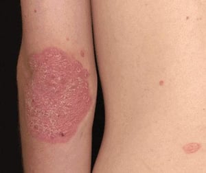
Photo provided by Thomas Habif, MD.

Photo provided by Thomas Habif, MD.

Image provided by Thomas Habif, MD.
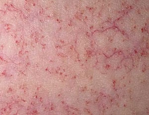
Photo provided by Thomas Habif, MD.

Photo courtesy of the Public Health Image Library of the Centers for Disease Control and Prevention.

Photo provided by Thomas Habif, MD.

Image provided by Thomas Habif, MD.


Photo provided by Thomas Habif, MD.

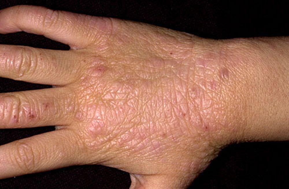
Photo provided by Thomas Habif, MD.

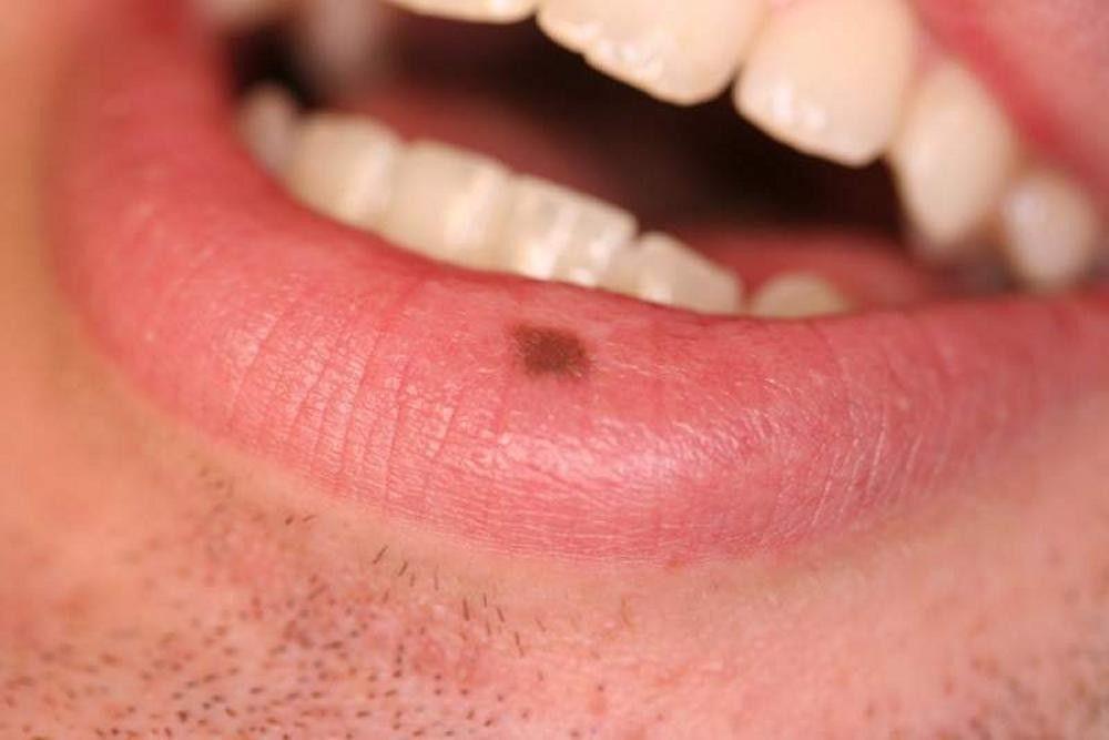
Photo provided by Robert MacNeal, MD.


Photo provided by Thomas Habif, MD.


Photo provided by Thomas Habif, MD.
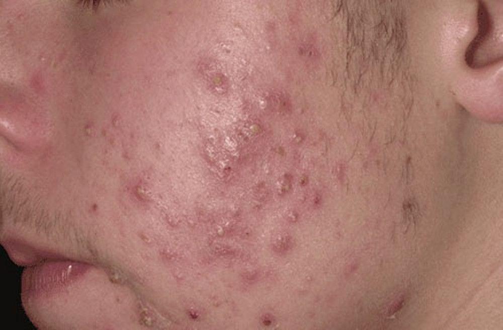
Photo provided by Thomas Habif, MD.

Image provided by Thomas Habif, MD.

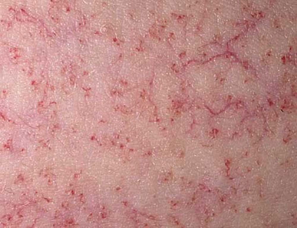
Photo provided by Thomas Habif, MD.

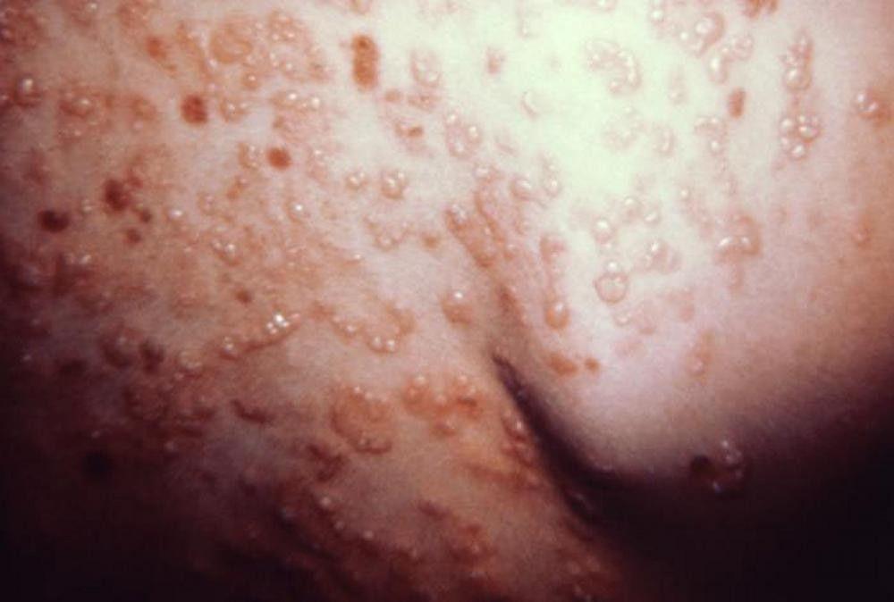
Photo courtesy of the Public Health Image Library of the Centers for Disease Control and Prevention.

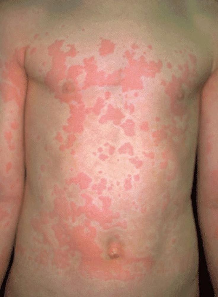
Photo provided by Thomas Habif, MD.

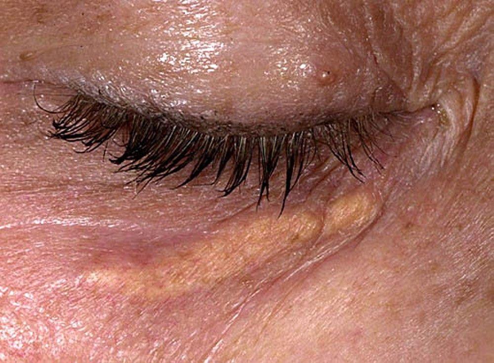
Image provided by Thomas Habif, MD.
Color Changes in the Skin
Although certain color changes are typical, the natural color of a person's skin can change the appearance of these colors.
Red skin (erythema) can result from many different disorders that cause inflammation or are caused by infection. Tumors on the skin are often pink or red, and disorders affecting blood vessels near the skin surface, such as port-wine stains, may appear red. In people who have dark skin, the same conditions that cause redness can be more subtle (with possibly less contrast with surrounding skin) and harder to recognize.
Orange skin is most often the result of hypercarotenemia. Hypercarotenemia is a condition that is the result of too much of the pigment carotene in the blood. People who overeat foods rich in beta-carotene, such as carrots, may develop hypercarotenemia. In people who have dark skin, the orange may be more noticeable on the palms and soles.
Yellow skin may occur in people who have jaundice. In people who have dark skin, the yellow may be more noticeable in the sclera (white part of the eye) than the skin. Causes of isolated yellow areas include xanthelasmas and xanthomas (small yellow deposits of fat in the skin or tendons) and pseudoxanthoma elasticum.
Green fingernails are typically caused by infection with the bacteria Pseudomonas aeruginosa.
Violet/purple skin may be caused by bleeding beneath the skin (cutaneous hemorrhage) or vasculitis. Abnormal overgrowths of blood vessels, such as Kaposi sarcoma and hemangiomas, can appear purple. Skin inflammation due to dermatomyositis may cause a reddish purple or lilac color around the eyes and face (called a heliotrope rash).
Shades of blue, silver, and graymoles (nevi) that are deep in the skin may appear blue.
Black skin lesions may contain specialized cells that produce the brown pigment melanin (melanocytes). Examples of these types of lesions include moles (nevi) and melanoma. Thick, black, crusty scabs (called eschars) are collections of dead skin and can be caused by death of tissue (infarction).


