


Mucormycosis is a fungal infection that can be caused by many different molds.
Mucormycosis is acquired when spores produced by the mold are inhaled or, much less commonly, when they enter the body through a cut or other break in the skin.
The infection causes pain, fever, and sometimes cough and can destroy structures in the face.
Doctors diagnose the infection by identifying the fungus in tissue samples.
Most people are given high doses of amphotericin B intravenously, and surgery is done to remove infected and dead tissue.
(See also Overview of Fungal Infections.)
Many different species of fungi can cause mucormycosis. They belong to a large group of molds called Mucorales. These molds include Rhizopus, Rhizomucor, and Mucor. Each species causes similar symptoms.
These molds are common in the environment and include many common bread molds. People probably breathe in the spores of these molds all the time. However, these molds usually do not cause infection.
Mucormycosis typically occurs when one of the following is present:
Diabetes is not controlled well.
The immune system is weakened by medications (such as corticosteroids or medications that suppress the immune system) or by leukemia or other disorders that weaken the immune system.
Mucormycosis can be caused by
Inhaling spores produced by the molds (the most common cause)
Having the mold's spores enter through a break in the skin
Inhaling the spores can cause several types of infection:
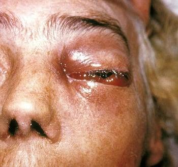
The nose, sinuses, eyes, and brain are most often infected. This severe infection—called rhinocerebral mucormycosis—is often fatal.
Spores can enter the lungs, causing pulmonary mucormycosis.
When spores inhaled into the mouth are swallowed, the digestive tract can be infected.
When the infection results from spores entering through a break in the skin, it affects the skin—a form called cutaneous mucormycosis. This form usually occurs in people with a normal immune system when contaminated soil comes in contact with broken skin, as may occur during earthquakes or other natural disasters or in people injured in blast injuries during combat.
Mucormycosis does not spread from person to person.
Symptoms of Mucormycosis
Rhinocerebral mucormycosis may cause pain, fever, sinus pain, and, if the eye socket is infected (called orbital cellulitis), bulging of the affected eye (proptosis). Vision may be lost.
The roof of the mouth (palate), the facial bones surrounding the eye socket or sinuses, or the divider between the nostrils (septum) may be destroyed by the infection. The dead tissue turns black.
Infection in the brain may cause difficulty using and understanding language, seizures, partial paralysis, and coma.

Pulmonary mucormycosis causes severe symptoms, including high fever, cough, and difficulty breathing.
In cutaneous mucormycosis, the area around the break in the skin may be warm, red, swollen, and painful. People may have a fever. Ulcers or blisters may form, and the infected tissue may turn black.
The fungus tends to invade arteries. As a result, blood clots form in the arteries, blood flow to tissues is blocked, and tissue dies. The fungus grows uncontrolled in the dead tissue, which turns black. The surrounding area may bleed.
Diagnosis of Mucormycosis
Culture and examination of samples of infected tissue
Because symptoms of mucormycosis can resemble those of other infections, a doctor may not be able to diagnose it immediately.
To diagnose mucormycosis, a doctor takes samples of infected tissue and sends them to a laboratory to be grown (cultured) and examined under a microscope. The diagnosis is made when the fungus is identified in the samples. However, sometimes these tests do not detect the fungus.
Doctors may do computed tomography (CT) or x-rays to check for damage to facial structures.
Blood tests may be done to gather information about the DNA of the fungus, which helps doctors identify it.
Did You Know...
|
Treatment of Mucormycosis
Antifungal medications
Treatment of the underlying condition
Surgery to remove infected and dead tissue
Early diagnosis and treatment of mucormycosis are important to prevent death or to avoid extensive surgery, which often causes disfigurement. Thus, treatment is started as soon as this infection is diagnosed or suspected.
Infected tissue and especially dead tissue must be removed by surgery.
Prognosis for Mucormycosis
Mucormycosis is very serious. Even when as much infected and dead tissue as possible is removed and antifungal medications are used appropriately, many people with this infection die.


