


Celiac disease is an immunologically mediated disease in genetically susceptible people caused by intolerance to gluten, resulting in mucosal inflammation and villous atrophy, which causes malabsorption. Symptoms usually include diarrhea and abdominal discomfort. Diagnosis is by small-bowel biopsies showing characteristic though not specific pathologic changes of villous atrophy that resolve with a strict gluten-free diet.
Celiac disease is a malabsorption disorder.
Etiology of Celiac Disease
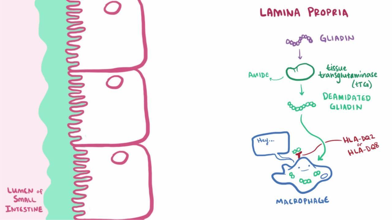
Celiac disease is a hereditary disorder caused by sensitivity to the gliadin fraction of gluten, a protein found in wheat; similar proteins are present in rye and barley. In a genetically susceptible person, gluten-sensitive T cells are activated when gluten-derived peptide epitopes are presented. The inflammatory response causes characteristic mucosal villous atrophy in the small bowel.
Epidemiology of Celiac Disease
Celiac disease mainly affects people of northern European descent. Prevalence estimates based on serologic screens among blood donors (sometimes confirmed by biopsy) indicate the disorder may be present in about 1/150 in Europe, especially in Ireland and Italy, and perhaps 1/250 in some parts of the United States. Current prevalence estimates in some regions are as high as 1/100.
The disease affects about 10 to 20% of 1st-degree relatives. Female:male ratio is 2:1. Onset is generally in childhood but may occur later.
Patients who have other diseases, such as lymphocytic colitis, Down syndrome, type 1 diabetes mellitus, and autoimmune (Hashimoto) thyroiditis, are at risk of developing celiac disease.
Symptoms and Signs of Celiac Disease
The clinical presentation varies; no typical presentation exists. Some patients are asymptomatic or have only signs of nutritional deficiency. Others have significant gastrointestinal symptoms.
Celiac disease can manifest in infancy and childhood after introduction of cereals into the diet. The child has failure to thrive, apathy, anorexia, pallor, generalized hypotonia, abdominal distention, and muscle wasting. Stools are soft, bulky, clay-colored, and foul-smelling. Older children may present with anemia or failure to grow normally.
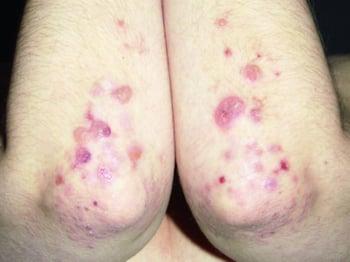
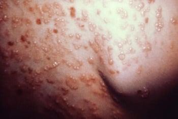
About 10% of patients have dermatitis herpetiformis, an intensely pruritic papulovesicular rash that is symmetrically distributed over the extensor areas of the elbows, knees, buttocks, shoulders, and scalp. This rash can be induced by a high-gluten diet.


Diagnosis of Celiac Disease
Serologic markers
Small-bowel biopsy
(See also the American College of Gastroenterology's 2023 Guidelines Update: Diagnosis and Management of Celiac Disease.)
The diagnosis of celiac disease is suspected clinically and by laboratory abnormalities suggestive of malabsorption. Family incidence is a valuable clue. Celiac disease should be strongly considered in a patient with iron deficiency without obvious gastrointestinal bleeding.
Confirmation requires a small-bowel biopsy from the second portion of the duodenum. Findings include lack or shortening of villi (villous atrophy), increased intraepithelial cells, and crypt hyperplasia. However, such findings can also occur in tropical sprue, severe small intestinal bacterial overgrowth, eosinophilic enteritis, infectious enteritis (eg, giardiasis), and lymphoma.
Because biopsy lacks specificity, serologic markers can aid diagnosis. Anti-tissue transglutaminase antibody (tTG) and anti-endomysial antibody (EMA—an antibody against an intestinal connective tissue protein) have sensitivity and specificity > 90%. These markers can also be used to screen populations with high prevalence of celiac disease, including 1st-degree relatives of affected patients and patients with diseases that occur at a greater frequency in association with celiac disease. If either test is positive, the patient should have a diagnostic small-bowel biopsy. If both are negative, celiac disease is extremely unlikely. These antibodies decrease in titer in patients on a gluten-free diet and thus are useful in monitoring dietary adherence. All diagnostic serologic testing should be done with patients following a gluten-containing diet.
Histocompatibility testing can be useful in selected clinical situations. More than 95% of celiac patients have the human leukocyte antigen (HLA)-DQ2 or HLA-DQ8 haplotype (1), although these haplotypes are not particularly specific for celiac disease. However, given the high sensitivity, testing that fails to show HLA-DQ2 or -DQ8 can effectively rule out celiac disease when biopsy and serologic markers are not concordant.
Malabsorption tests are not specific for celiac disease. If done, common findings include steatorrhea of 10 to 40 g/day and abnormal results with D-xylose and (in severe ileal disease) positive Schilling tests.
Pearls & Pitfalls
|
Diagnosis reference
1. Kaukinen K, Partanen J, Mäki M, Collin P: HLA-DQ typing in the diagnosis of celiac disease. Am J Gastroenterol 97(3):695–699, 2002. doi: 10.1111/j.1572-0241.2002.05471.x
Treatment of Celiac Disease
Gluten-free diet
Supplements to replace any serious deficiencies
(See also the American College of Gastroenterology's 2023 Guidelines Update: Diagnosis and Management of Celiac Disease.)
Treatment of celiac disease is a gluten-free diet (avoiding foods containing wheat, rye, or barley). Gluten is so widely used (eg, in commercial soups, sauces, ice creams, and hot dogs) that a patient needs a detailed list of foods to avoid. Patients are encouraged to consult a dietitian and join a celiac support group such as Beyond Celiac or the Celiac Disease Foundation. The response to a gluten-free diet is usually rapid, and symptoms resolve in 1 to 2 weeks. Ingesting even small amounts of food containing gluten may prevent remission or induce relapse.
Small-bowel biopsy should be repeated after 3 to 6 months of a gluten-free diet. If abnormalities persist, other causes of villous atrophy (eg, lymphoma) should be considered. Lessening of symptoms and improvement in small-bowel morphology are accompanied by a decrease in anti-tissue transglutaminase antibody and anti-endomysial antibody titers.
Supplementary vitamins, minerals, and hematinics may be given, depending on the deficiencies. Mild cases may not require supplementation, whereas severe cases may require comprehensive replacement. For adults, replacement includes oral ferrous sulfate 300 mg once every other day to 3 times a day, oral folate 5 to 10 mg once/day, calcium supplements, and any standard multivitamin. Sometimes children (but rarely adults) who are seriously ill on initial diagnosis require bowel rest and total parenteral nutrition.
If a patient responds poorly to gluten withdrawal, either the diagnosis is incorrect or the disease has become refractory. Corticosteroids can control symptoms in refractory disease.
Prognosis for Celiac Disease
Complications of celiac disease include refractory disease, collagenous sprue, and intestinal lymphomas.
Intestinal lymphomas affect 6 to 8% of patients with celiac disease, usually manifesting after 20 to 40 years of disease. The incidence of other gastrointestinal cancers (eg, carcinoma of the esophagus or oropharynx, small-bowel adenocarcinoma) also increases (1). Adherence to a gluten-free diet can significantly reduce the risk of cancer.
If people who have been doing well on a gluten-free diet for a long time once again develop symptoms of celiac disease, physicians usually do upper endoscopy with small-bowel biopsy and/or capsule endoscopy to check for signs of intestinal lymphoma.
Prognosis reference
1. Ilus T, Kaukinen K, Virta LJ, et al: Incidence of malignancies in diagnosed celiac patients: A population-based estimate. Am J Gastroenterol 109(9):1471–1477, 2014. doi: 10.1038/ajg.2014.194
Key Points
Celiac disease involves an inflammatory response to gluten that causes villous atrophy and malabsorption.
People of northern European heritage are most often affected.
Suspect the diagnosis if the serologic markers anti-tissue transglutaminase antibody and anti-endomysial antibody are present and confirm the diagnosis with a small-bowel biopsy.
Instruct the patient to follow a gluten-free diet and replace any vitamin or mineral deficiencies.
More Information
The following English-language resources may be useful. Please note that THE MANUAL is not responsible for the content of these resources.
American College of Gastroenterology: Guidelines Update: Diagnosis and Management of Celiac Disease (2023)
For patients, the following sites provide information about celiac disease, including how to live with it, what to eat, treatment options, and clinical trials:


