


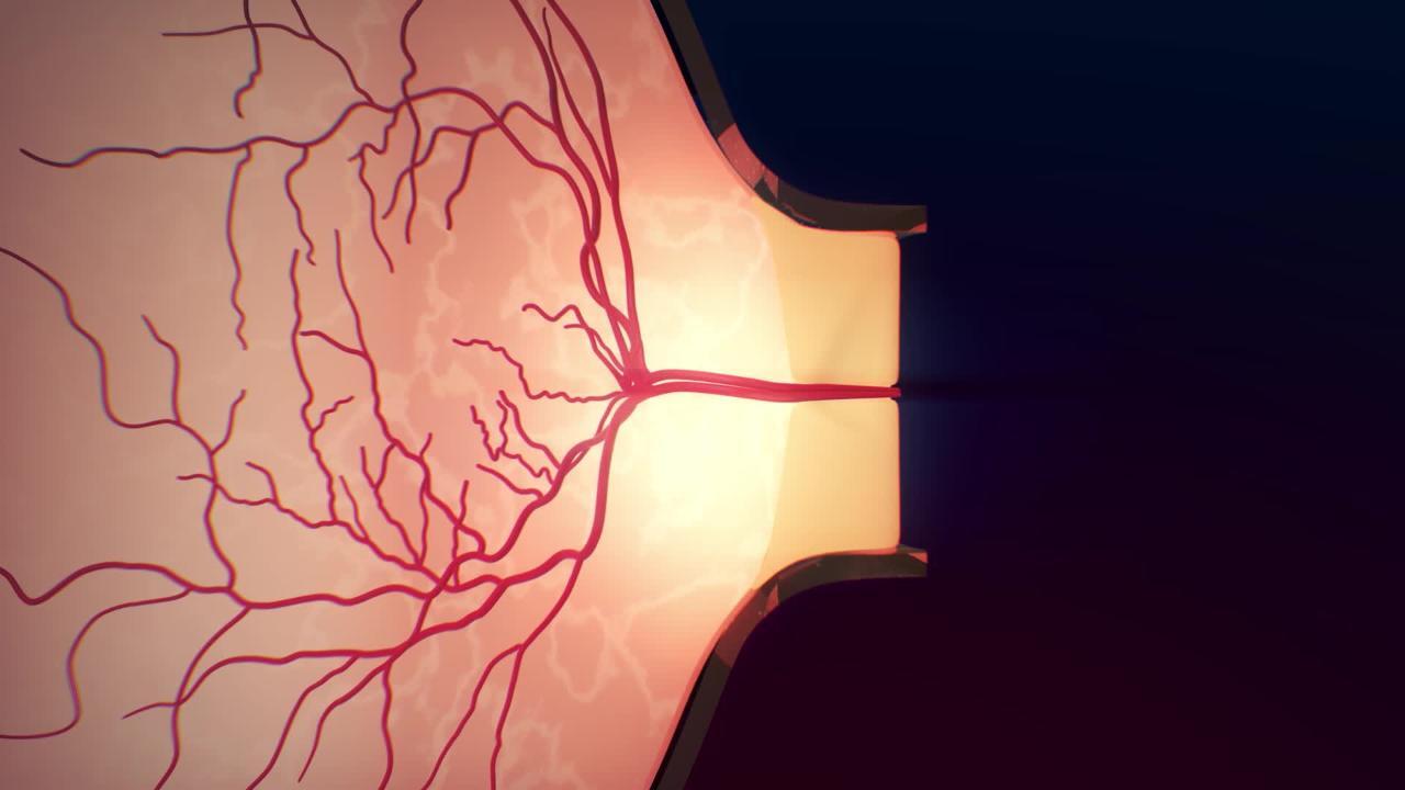
Papilledema is a condition in which increased pressure in or around the brain causes the part of the optic nerve inside the eye to swell.
Visual symptoms may be fleeting disturbances in vision.
Other symptoms of increased pressure in or around the brain include whooshing noise in the ears, headache, vomiting, or a combination.
Doctors make the diagnosis by looking in the person’s eye with an ophthalmoscope.
The disorder causing increased brain pressure is treated as soon as possible.
(See also Overview of Optic Nerve Disorders.)
Causes of Papilledema
Papilledema is usually caused by the following:
Idiopathic intracranial hypertension (most common cause)
Bleeding in the brain
Inflammation of the brain (encephalitis) or its tissue coverings (meningitis)
Uncontrolled, life-threatening hypertension
A blood clot in parts of some large veins within the brain (cerebral venous sinus thrombosis)
These conditions typically result in papilledema in both eyes.
Symptoms of Papilledema
At first, papilledema may be present without affecting vision. Fleeting vision changes—blurred vision, double vision, flickering, or complete loss of vision—typically lasting seconds are characteristic of papilledema. Other symptoms may be caused by the elevated pressure in the brain. A pulsating whooshing noise in the ears, headache, nausea, vomiting, or a combination may occur. This disorder does not cause eye pain.
Diagnosis of Papilledema
A doctor's evaluation
Imaging tests
Lumbar puncture (spinal tap)
To diagnose papilledema, a doctor uses an ophthalmoscope (a light with magnifying lenses that is used to look into the back of the eye). Often an ophthalmologist (a medical doctor who specializes in the evaluation and treatment of eye disorders) needs to confirm the diagnosis and help determine the cause.
Magnetic resonance imaging (MRI) or computed tomography (CT) of the brain and orbits may be used to help determine the cause and monitor the effect of treatment. MR venogram or CT venogram of the head can be done to rule out a cerebral venous sinus thrombosis.
A lumbar puncture (spinal tap) is done to measure the pressure of the cerebrospinal fluid unless something is seen on the MRI or CT scan indicating a spinal tap is not safe to do. A sample of the cerebrospinal fluid may be examined for evidence of a brain tumor or infection.
Sometimes ultrasonography of the eye is done to distinguish between papilledema and other disorders that cause apparent swelling of the optic nerve. Optical coherence tomography (OCT) is a specialized technique that uses reflected light to create a more detailed image of the back of the eye and optic nerve.
Treatment of Papilledema
Treatment of cause
The disorder causing increased brain pressure is treated as soon as possible. For example, if the high pressure of the cerebrospinal fluid is caused by a brain tumor, corticosteroids may be given, but surgery to remove the tumor or radiation therapy to decrease its size may be needed.
Papilledema that occurs as a result of idiopathic intracranial hypertension can be treated with weight loss and a diuretic. If unsuccessful, surgical procedures can be done.
An infection, if bacterial, can be treated with antibiotics.
A brain abscess is drained, and antibiotics are given.


