


Tumors that originally start in the bone are called primary bone tumors. Primary bone tumors may be noncancerous (benign) or cancerous (malignant).
After cancer is diagnosed, it is staged. Staging is a way of describing how advanced the cancer has become, including such criteria as degree of aggressiveness (how likely it is to spread, based on how the tumor cells appear under a microscope), how big it is, and whether it has spread to neighboring tissues or more distantly to lymph nodes or other organs.
(See also Overview of Bone Tumors and Overview of Cancer.)
Adamantinomas
Adamantinomas are rare tumors that most often develop in the shinbone (tibia). The tumors usually occur in adolescents and people who are in their 20s but can occur at any age. They often cause pain, and people often can feel the tumor beneath the skin when they run their fingers over it.
These tumors grow slowly and are low-grade cancers, which means they are less likely to spread (metastasize) than some other tumors. However, although rare, metastases do occur (mostly to the lungs).
To diagnose adamantinomas, doctors take x-rays and remove a tissue sample for examination under a microscope (biopsy).
To treat adamantinomas, doctors surgically remove them without cutting into the tumor, which risks spilling the tumor cells. If the cells spill, the cancer can return. On rare occasions, surgical removal of the affected leg (amputation) may be necessary depending on the location of the tumor or depending on whether the tumor returns.
Chondrosarcomas
Chondrosarcomas are tumors composed of cancerous cartilage cells. These tumors tend to occur in older adults. These tumors often develop in bones such as the pelvis or shoulder blade (scapula) but can develop in any portion of any bone and can also develop in the tissues surrounding the bones. Many chondrosarcomas are slow-growing or low-grade tumors, meaning that they are less likely to spread (metastasize) than some other tumors. However, some chondrosarcomas are fast-growing or high-grade tumors, which tend to metastasize.
To diagnose chondrosarcomas, doctors take x-rays and do a bone scan and magnetic resonance imaging (MRI). Doctors also remove a tissue sample for examination under a microscope (biopsy).
High-grade or fast-growing chondrosarcomas are aggressive tumors and are more likely to metastasize than some other tumors. They must be completely removed surgically without cutting into the tumor, which risks spilling the tumor cells. If the cells spill, the cancer can return.
Chondrosarcomas of any grade do not respond to chemotherapy or radiation therapy. Surgical removal of the affected arm or leg (amputation) is rarely necessary.
Chordomas
Chordomas are rare and cancerous and tend to occur at the ends of the spinal column, usually in the middle of the base of the spine (sacrum) or tailbone or near the base of the skull. A chordoma affecting the sacrum or tailbone causes nearly constant pain. A chordoma in the base of the skull can cause problems in nerves at the base of the skull (the cranial nerves). Symptoms may exist for months to several years before diagnosis. Chordomas do not usually spread (metastasize) to other areas such as the lung unless they are more aggressive, but they may return after treatment.
To help diagnose chordomas, doctors do magnetic resonance imaging (MRI). Doctors also do a biopsy.
Chordomas affecting the sacrum or tailbone may be cured by surgical removal. Chordomas in the base of the skull usually cannot be cured surgically, but radiation therapy may temporarily shrink the tumor and help with pain.
Ewing sarcoma of bone
Ewing sarcoma is a cancerous tumor that affects males more often than females and appears most commonly in people aged 10 to 20 years. Most of these tumors develop in the arms or legs, but they may develop in any bone. Pain and swelling are the most common symptoms. Tumors may become quite large, sometimes affecting the entire length of a bone. The tumor may include a large mass of soft tissue.
To diagnose Ewing sarcoma, doctors take x-rays. Although x-rays can show some details, magnetic resonance imaging (MRI) can help determine the exact size of the tumor. To confirm the diagnosis, doctors do a biopsy.

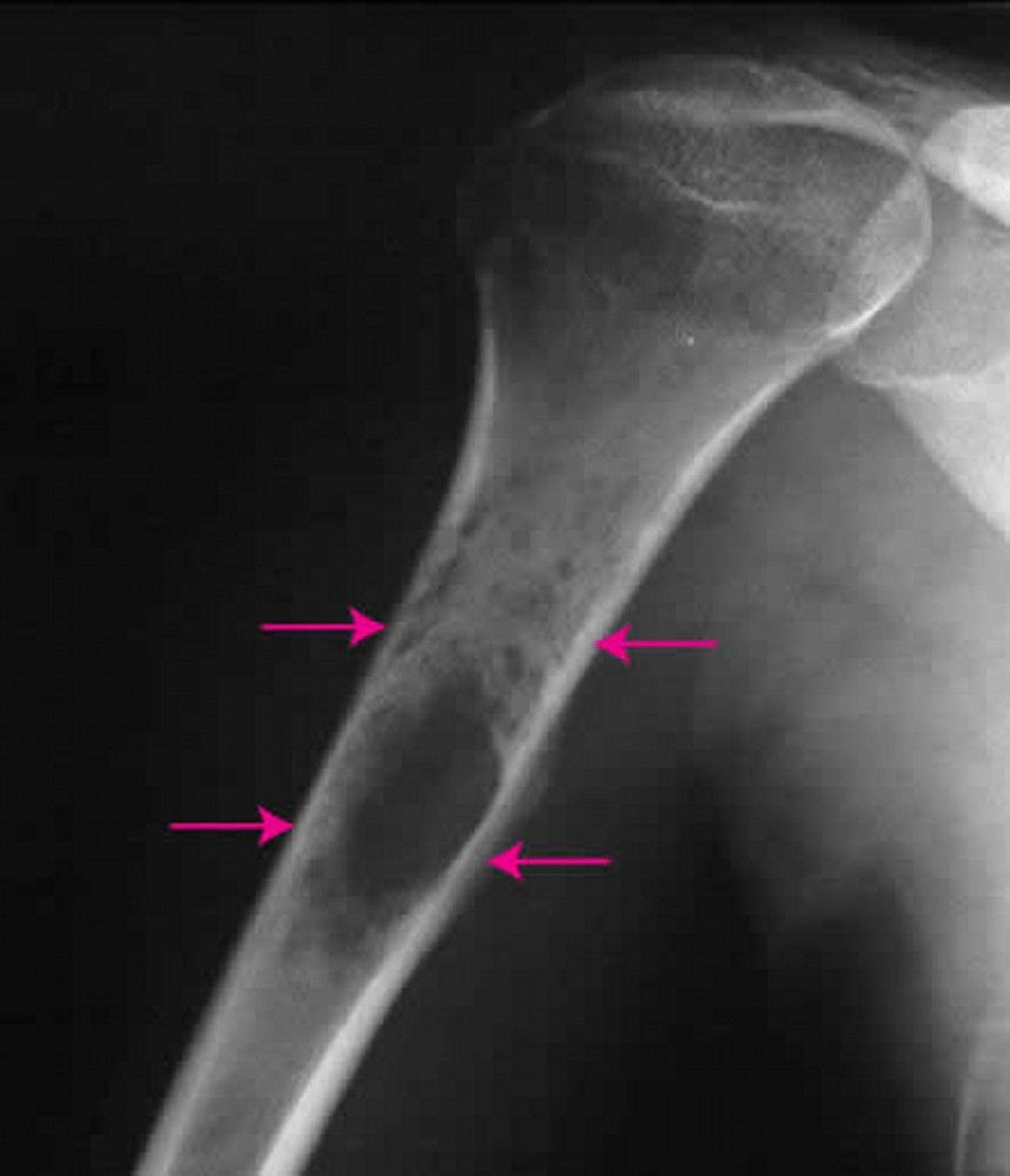
Image courtesy of Michael J. Joyce, MD, and Hakan Ilaslan, MD.
Treatment of Ewing sarcoma includes various combinations of surgery, chemotherapy, and radiation therapy, depending on whether surgery is practical or, if attempted, successful. These treatment combinations can cure more than 60% of people who have Ewing sarcoma.
Fibrosarcomas and undifferentiated pleomorphic sarcomas of bone
Fibrosarcomas and undifferentiated pleomorphic sarcomas of bone (formerly known as malignant fibrous histiocytoma of bone) affect the same age group as and are similar to osteosarcomas in appearance, location, and symptoms. These cancerous tumors have cells that produce cancerous fibrous tissue (connective tissue) rather than cancerous bony tissue.
Treatment and survival rates are similar to that of osteosarcoma.
Lymphoma of bone
Lymphoma of bone (previously called reticulum cell sarcoma) is a cancerous tumor that usually affects people in their 40s and 50s. It can originate in any bone or elsewhere in the body and then spread diffusely to bone marrow. Usually, this tumor causes pain and swelling and an accumulation of soft tissue. The damaged bone tends to break (fracture).
To diagnose lymphoma of bone, doctors take x-rays and do magnetic resonance imaging (MRI). Biopsy is also done.
Treatment of lymphoma of bone usually consists of a combination of chemotherapy with or without radiation therapy, which seems to be as effective as surgical removal of the tumor. Amputation is rarely necessary. If a bone seems as though it may fracture, doctors may stabilize it surgically in an attempt to prevent a fracture.
Malignant giant cell tumors
Malignant giant cell tumors are rare and cancerous and are usually located at the extreme end of a long bone (arm or thigh bone). These typically cause pain and swelling.
To diagnose malignant giant cell tumors, doctors take x-rays and also do magnetic resonance imaging (MRI) and a biopsy.
Treatment of malignant giant cell tumors is similar to that of osteosarcomas, but the cure rate is low.
Multiple myeloma
Multiple myeloma (see also Plasma Cell Disorders: Multiple Myeloma) is sometimes considered a cancer of the hematologic (blood) system, but is sometimes considered a tumor of bone. As a bone tumor, it is the most common primary cancerous (malignant) bone tumor and occurs mostly in older adults. However, it is cancer that involves the bone marrow (the blood-forming tissue inside the cavity of the bone) rather than the hard tissue that makes up the bone. Thus, it is usually considered a cancer of the bone marrow rather than the bone itself. It is more common than cancers of the hard tissue that makes up bone.
The cancerous marrow cells secrete substances that cause loss of bone. Bone loss may be widespread or, more often, appears as punched-out areas in bones.
Multiple myeloma may affect one or more bones, so pain may occur in one location or in several. If only one bone is affected by a single tumor, the condition is called plasmacytoma. If there is more than one tumor or the bone marrow is widely affected, the condition is called multiple myeloma.
Bone biopsy is done sometimes for diagnosis in areas where bone has been destroyed. If multiple myeloma is suggested by the bone biopsy results or if multiple myeloma is suspected for other reasons, the diagnosis is confirmed by removing and examining bone marrow cells. Blood tests are also done. Additionally, doctors take x-rays of the entire body (skeletal survey). Magnetic resonance imaging (MRI) or positron emission tomography (PET) combined with computed tomography (PET-CT) may also be done to examine specific sites of bone pain.

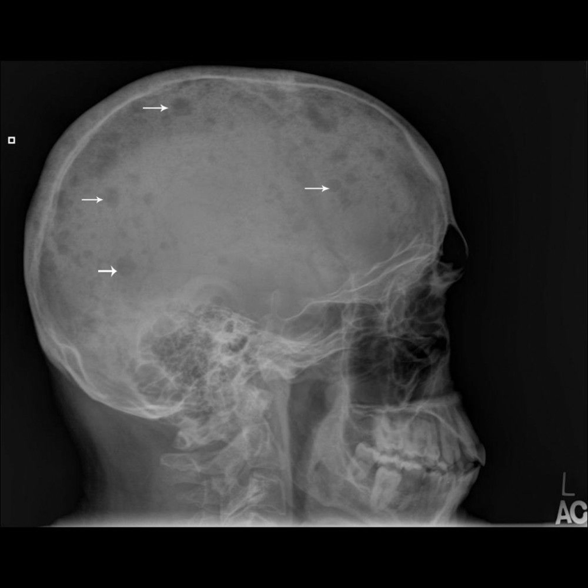
Image courtesy of Michael J. Joyce, MD, and Hakan Ilaslan, MD.
Treatment of multiple myeloma is complex and may include chemotherapy, radiation therapy, and sometimes surgery.
Osteosarcomas (osteogenic sarcoma)
Osteosarcoma is the most common type of primary cancerous bone tumor if one considers multiple myeloma as a hematologic tumor. Although most common among people aged 10 to 25 years, osteosarcomas can occur at any age. There is a genetic predisposition, especially in children who carry the gene for hereditary retinoblastoma and Li-Fraumeni syndrome. Older people who have Paget disease of bone, have undergone bone radiation therapy for another cancer, or have areas of dead bone tissue (called bone infarcts) and other conditions sometimes develop this type of tumor. Osteosarcomas usually develop in or around the knee, but they can originate in any bone. They tend to spread (metastasize) to the lungs or other bones. Usually, these tumors cause pain and swelling.
X-rays are taken, but removal of a tissue sample for examination under a microscope (biopsy) is needed for the diagnosis of osteosarcoma. People need a chest x-ray and a chest computed tomography (CT) scan to detect cancer that has metastasized to the lungs and a bone scan to detect cancer that has spread to other bones. Magnetic resonance imaging (MRI) and positron emission tomography (PET) combined with computed tomography (PET-CT) are other imaging tests that are also done.

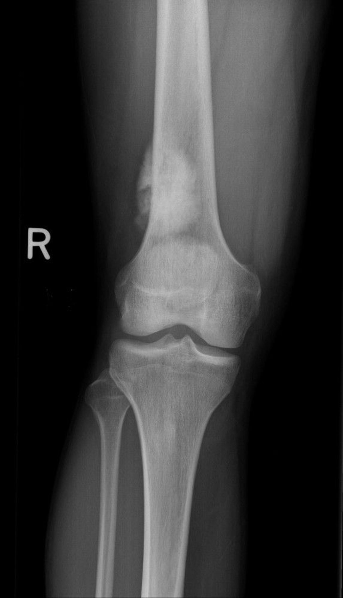
Image courtesy of Michael J. Joyce, MD, and Hakan Ilaslan, MD.
More than 65% of people who have this type of tumor survive for at least 5 years after diagnosis when chemotherapy is given and the cancer has not metastasized. If chemotherapy destroys almost all of the cancer, the chance of surviving at least 5 years is greater than 90%. Because surgical procedures have improved, the affected arm or leg can usually be saved and reconstructed. In the past, the affected limb often had to be amputated.
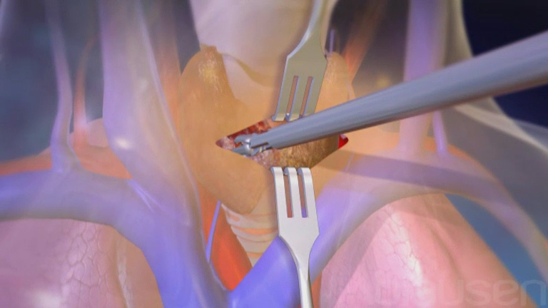
Osteosarcomas are usually treated with a combination of chemotherapy and surgery. Usually, chemotherapy is given first. Pain often subsides during this phase of treatment. Then the tumor is surgically removed without cutting into the tumor. Cutting into the tumor spills its cells, which can cause the cancer to return in the same area. Chemotherapy continues after surgery.


