


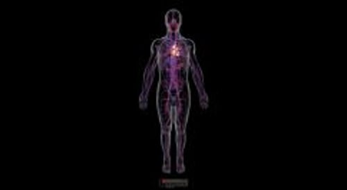
The heart and blood vessels constitute the cardiovascular (circulatory) system. The heart pumps the blood to the lungs so it can pick up oxygen and then pumps oxygen-rich blood to the body. The blood circulating in this system delivers oxygen and nutrients to the tissues of the body and removes waste products (such as carbon dioxide) from the tissues.
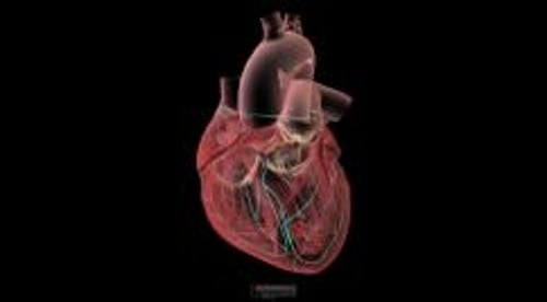
The heart, a hollow muscular organ, is located in the center of the chest. The heart has two sides, right and left. The right and left sides of the heart each have an
Atrium: Upper chamber that collects blood and pumps it to the lower chamber
Ventricle: Lower chamber, which pumps blood out of the heart
To ensure that blood flows in only one direction, each ventricle has an "in" (inlet) valve and an "out" (outlet) valve.
In the left ventricle, the inlet valve is the mitral valve, and the outlet valve is the aortic valve. In the right ventricle, the inlet valve is the tricuspid valve, and the outlet valve is the pulmonic (pulmonary) valve.
Each valve consists of flaps (cusps or leaflets), which open and close like one-way swinging doors. The mitral valve has two cusps. The other valves (tricuspid, aortic, and pulmonic) have three. The large inlet valves (mitral and tricuspid) have tethers—consisting of the papillary muscles and cords of tissue—which prevent the valves from swinging backward into the atria. If a papillary muscle is damaged (for example, by a heart attack), the valve may then swing backward and start leaking (called regurgitation). If a valve opening is narrowed (called stenosis), blood flow through the valve is reduced. Both leaking and narrowing may occur in the same valve.
The heartbeats are evidence that the heart is pumping. Doctors often describe the sound of the heartbeat as lub-dub. When doctors listen to the heartbeat with a stethoscope, the first sound they hear (the lub of lub-dub) is the sound of the mitral and tricuspid valves closing. The second sound (the dub) is the sound of the aortic and pulmonic valves closing. Each heartbeat has two parts:
Systole: During systole, the ventricles contract and pump blood out of the heart, and the atria relax and begin filling with blood again.
Diastole: During diastole, the ventricles relax and fill with blood. Then the atria contract, forcing more blood into the ventricles.
Function of the Heart
The heart's only function is to pump blood.
The right side of the heart: Pumps blood to the lungs, where oxygen is added to the blood and carbon dioxide is removed
The left side of the heart: Pumps blood to the rest of the body, where oxygen and nutrients are delivered to tissues and waste products (such as carbon dioxide) are transferred to the blood for removal by other organs (such as the lungs and kidneys)
A Look Into the Heart
This cross-sectional view of the heart shows the direction of normal blood flow. |

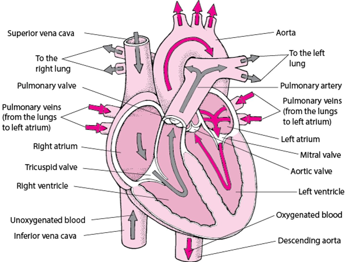
Blood travels the following circuit: Blood from the body, which is depleted of oxygen and laden with carbon dioxide, flows through the two largest veins—the superior vena cava and the inferior vena cava, known collectively as the venae cavae—into the right atrium. When the right ventricle relaxes, blood in the right atrium pours through the tricuspid valve into the right ventricle. When the right ventricle is nearly full, the right atrium contracts, propelling additional blood into the right ventricle, which then contracts. This contraction closes the tricuspid valve and propels blood through the pulmonary valve into the pulmonary arteries, which supply the lungs. In the lungs, blood flows through the tiny capillaries that surround the air sacs. Here, the blood absorbs oxygen and gives up carbon dioxide, which is then exhaled.
Blood from the lungs, which is now oxygen-rich, flows through the pulmonary veins into the left atrium. When the left ventricle relaxes, the blood in the left atrium pours through the mitral valve into the left ventricle. When the left ventricle is nearly full, the left atrium contracts, propelling additional blood into the left ventricle, which then contracts. (In older people, the left ventricle does not fill as well before the left atrium contracts, making this contraction of the left atrium especially important.) The contraction of the left ventricle closes the mitral valve and propels blood through the aortic valve into the aorta, the largest artery in the body. This blood carries oxygen to all of the body except to the lungs.
The pulmonary circulation is the circuit through the right side of the heart, the lungs, and the left atrium.
The systemic circulation is the circuit through the left side of the heart, most of the body, and the right atrium.
Blood Supply of the Heart
Like all organs, the heart needs a constant supply of oxygen-rich blood. Even though the heart chambers are full of blood, the heart muscle needs its own dedicated blood supply which is called the
Coronary circulation
The coronary circulation is the system of arteries and veins that supplies the heart muscle (myocardium) with oxygen-rich blood and then returns oxygen-depleted blood to the right atrium.
The right coronary artery and the left coronary artery branch off the aorta (just after it leaves the heart) to deliver oxygen-rich blood to the heart muscle. These two arteries branch into other arteries that also supply blood to the heart. The cardiac veins collect blood from the heart muscle and empty it into a large vein on the back surface of the heart called the coronary sinus, which returns the blood to the right atrium. Because of the great pressure exerted in the heart as it contracts, most blood flows through the coronary circulation only while the ventricles are relaxing between beats (during diastole).
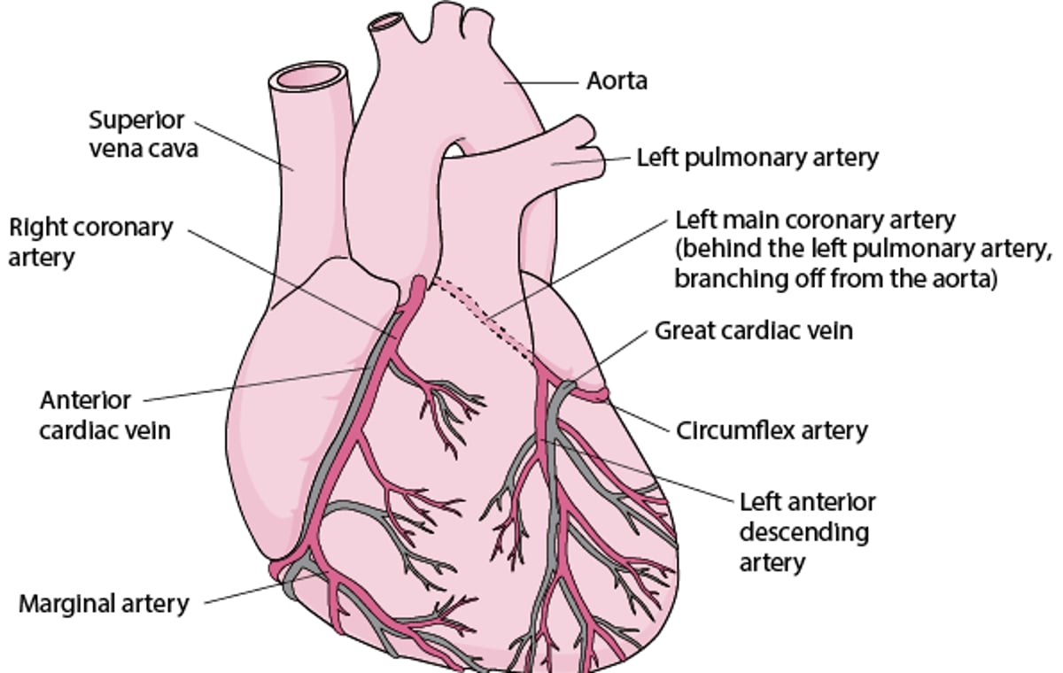
Supplying the Heart With Blood
Like any other tissue in the body, the muscle of the heart must receive oxygen-rich blood and have waste products removed by the blood. The right coronary artery and the left coronary artery, which branch off the aorta just after it leaves the heart, deliver oxygen-rich blood to the heart muscle. The right coronary artery branches into the marginal artery and the posterior interventricular artery, located on the back surface of the heart. The left coronary artery (typically called the left main coronary artery) branches into the circumflex and the left anterior descending artery. The cardiac veins collect blood containing waste products from the heart muscle and empty it into a large vein on the back surface of the heart called the coronary sinus, which returns the blood to the right atrium. |

Regulation of the Heart
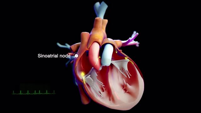
The contraction of the muscle fibers in the heart is very organized and highly controlled. Every heart muscle fiber does not contract at exactly the same time. Instead, the fibers contract in a sequence that best pumps blood out of each heart chamber. The contraction sequence is controlled by rhythmic electrical impulses (discharges) that flow through the heart in a precise manner along distinct pathways and at a controlled speed. The impulses originate in the heart's natural pacemaker (the sinus or sinoatrial node—a small mass of tissue in the wall of the right atrium), which generates a tiny electrical current.
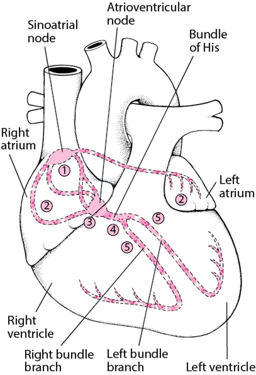
Tracing the Heart’s Electrical Pathway
The sinoatrial (sinus) node (1) initiates an electrical impulse that flows through the right and left atria (2), making them contract. When the electrical impulse reaches the atrioventricular node (3), it is delayed slightly. The impulse then travels down the bundle of His (4), which divides into the right bundle branch for the right ventricle (5) and the left bundle branch for the left ventricle (5). The impulse then spreads through the ventricles, making them contract. |

The heart rate, or pulse, is the number of times the heart beats within a minute. The heart rate goes up when the body needs more oxygen (such as during exercise). The heart rate goes down when the body needs less oxygen (such as during rest).
The rate at which the sinus node sends out its impulses governs the heart rate. The sinus node has its own rate of sending out impulses. This rate can be modified by two opposing parts of the autonomic nervous system—one part speeds the heart rate up (the sympathetic division of the nervous system) and one slows it down (the parasympathetic division).
The sympathetic division works through a network of nerves called the sympathetic plexus and through the hormones epinephrine (adrenaline) and norepinephrine (noradrenaline), which are released by the adrenal glands and the nerve endings.
The parasympathetic division works through a single nerve—the vagus nerve—which releases the neurotransmitter acetylcholine.


